Alzheimer's disease is characterized by the accumulation of amyloid β peptides and impaired glucose metabolism in the brain. Traditionally when there is impaired glucose metabolism the focus is on:

  • Sugar
  • Insulin
  • And cholesterol

Yet there are other biological entities at work in this case. Osteocalcin, an homonal protein, is secreted by osteoblasts. Osteoblasts are cells that synthesize bone. enter image description here 
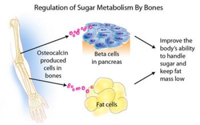
Patients with type 2 diabetes mellitus (T2DM) and/or cardiovascular disease (CVD), conditions of hyperinsulinaemia, have lower levels of osteocalcin and bone remodelling, and increased rates of fragility fractures.

In this study, daily intraperitoneal injection of osteocalcin for 4 weeks ameliorated the anxiety-like behaviors and cognitive dysfunctions in the APP/PS1 transgenic Alzheimer's disease mice model enter image description here Aβ burden in the hippocampus and cortex of Alzheimer's disease mice was ameliorated by osteocalcin. Besides, osteocalcin improved the neural network function of the brain, mainly in the enhanced power of high gamma band in the medial prefrontal cortex of Alzheimer's disease mice.

The proliferation of astrocytes in the hippocampus in Alzheimer's disease mice was also inhibited by osteocalcin Furthermore, OCN enhanced glycolysis in astrocytes and microglia.

Such an effect was abolished when the receptor of osteocalcin (Gpr158) was knockdown in astrocytes.

So osteocalcin maybe a novel therapeutic factor for Alzheimer's disease potentially through reducing Aβ burden and upregulation of glycolysis in neuroglia.

It should be noted that osteocalcin production is accelerated in acute stress response (like in physical effort), which stimulates osteocalcin release from bone within minutes humans. Vitamin D deficiency reduces osteocalcin synthesis and favours osteocyte apoptosis while its synthesis depends on vitamin K avilability.

Read the original article on Pubmed

It is generally accepted that dynamic changes resulting from the death of cells in the substantia nigra, a region of the midbrain, that lead to a dopamine deficit, underlie Parkinson's disease. enter image description here 
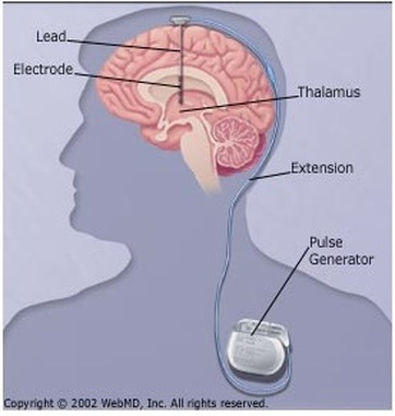
Deep brain stimulation of the subthalamic nucleus is a well-established treatment for the motor symptoms of Parkinson's disease, but it is still poorly understood how subthalamic stimulation modulates the state dynamics of the brain's motor network. A biological network represents systems as complex sets of relationships between various biological entities.

To investigate this, the authors of a new paper acquired functional resting-state magnetic resonance imaging time series data from 27 drug-free patients with Parkinson's disease who had brain stimulation electrodes. deep deployed in the subthalamic nucleus, in activated and deactivated states of stimulation. Sixteen matched healthy people were included as a control group.

In this paper, scientists adopted a modeling approach known as the hidden Markov model, to highlight the emergence of recurrent activation patterns of interacting motor regions via the oxygen level-dependent signal in blood detected in resting-state functional magnetic resonance of all participants. The hidden Markov model includes the dynamics of distinct states of the whole-brain motor network, including frequency of occurrence, duration, fractional coverage, and their transition probabilities between different states.

Notably, subthalamic stimulation reshapes network expression and stabilizes state transitions. In addition, subthalamic stimulation ameliorates motor symptoms by modulating transition trajectories between network states.

This modulating mechanism of subthalamic stimulation appears to have three significant effects: recovery, slowing, and remodeling effects. Significantly, recovery effects were correlated with improvement in subthalamic stimulation-induced tremor and postural symptoms.

Additionally, subthalamic stimulation was found to restore a relatively low level of functional connectivity fluctuation in all motor regions that is at a level close to that of healthy participants.

The authors' findings provide mechanistic insight and explanation of how subthalamic stimulation modulates motor symptoms in Parkinson's disease.

Read the original article on Pubmed

The risk of dementia is higher in women than in men. enter image description hereThe metabolic consequences of the decline in estrogen during menopause accelerate this neuropathology in women. However, the use of hormone replacement therapy gives contradictory results in the prevention of cognitive decline.

Hormone replacement therapy (HRT) is usually used to treat symptoms associated with female menopause. Within 10 years of menopause, hormone replacement therapy may reduces all-cause mortality and the risk of coronary heart disease, osteoporosis and dementia. But after 10 years, the beneficial effects on mortality and coronary heart disease are no longer apparent.

In this new publication, the authors study the modulating role of APOE genotype and age at the start of hormone replacement therapy on the heterogeneity of the cognitive response to hormone replacement therapy.

The analysis used baseline data from participants in the European Alzheimer's Dementia Prevention Cohort. Covariate pattern analysis was used to test the impact of APOE genotype and hormone replacement therapy on certain cognitive tests, such as MMSE, RBANS, score counting, the Four Mountain test, and the of the supermarket trolley, as well as the volumes of the medial temporal lobe by MRI.

APOE4 hormone replacement therapy users had the highest RBANS Delayed Memory Index score compared to APOE4 HRT non-users and APOE4 non-carriers, with entorhinal and amygdala volumes 6-10% greater important. Earlier initiation of hormone replacement therapy was associated with greater right and left hippocampal volumes only in APOE4 carriers.

Thus the introduction of hormone replacement therapy seems to be associated with delayed memory improvement and greater brain volumes only in in APOE4 carriers.

This may represent an effective targeted strategy to mitigate the higher lifetime risk of Alzheimer's disease in this large at-risk population subgroup. Confirmation of the results of a fit-for-purpose RCT with prospective recruitment based on APOE genotype is needed to establish causation.

We should remember that some studies on hormone replacement therapy seem to show that the risks could outweigh the benefits. It has been suggested that many of the positive perceptions of hormone replacement therapy may be due to the efforts of pharmaceutical companies including by publishing biased scientific publications.

Read the original article on Pubmed

The direct link between neuropathology and the symptoms that emerge from damage to the brain is often difficult to discern. In this perspective, the authors of this new publication argue that a satisfying account of neurodegenerative symptoms most naturally emerges from the consideration of the brain from the systems-level. enter image description here Source.

Specifically, the authors will highlight the role of the neuromodulatory arousal system, which is uniquely positioned to coordinate the brain's ability to flexibly integrate the otherwise segregated structures required to support higher cognitive functions.

Neuromodulatory systems, including the noradrenergic, serotonergic, dopaminergic, and cholinergic systems, track environmental signals, such as risks, rewards, novelty, effort, and social cooperation. These systems provide a foundation for cognitive function in higher organisms; attention, emotion, goal-directed behavior, and decision-making derive from the interaction between the neuromodulatory systems and brain areas, such as the amygdala, frontal cortex, hippocampus, and sensory cortices.

The existence of extensive lines of communication between the nervous system and immune system represents a fundamental principle underlying neuroinflammation.

Important sources in the above neuropathologies appear to be microglia and mast cells, together with astrocytes and possibly also oligodendrocytes. Understanding neuroinflammation also requires an appreciation that non-neuronal cell—cell interactions, between both glia and mast cells and glia themselves, are an integral part of the inflammation process.

Given their strong influence on behavior and cognition, these systems also play a key role in disease states and are the primary target of many current treatment strategies. The fact that these systems interact with each other either directly or indirectly, however, makes it difficult to understand how a failure in one or more systems can lead to a particular symptom or pathology.

Importantly, the neuromodulatory arousal system is highly heterogeneous, encompassing structures that are common sites of neurodegeneration across Alzheimer's and Parkinson's disease.
The scientists here will review studies that implicate the dysfunctional interactions amongst distributed brain regions as a side-effect of pathological involvement of the neuromodulatory arousal system in these neurodegenerative disorders.

From this perspective, the authors will argue that future work in clinical neuroscience should attempt to consider the inherent complexity in the brain and employ analytic techniques that do not solely focus on regional functional impairments, but rather captures the brain as an inherently dynamic, distributed, multi-scale system.

Through this lens, the authors hope that the authors will devise new and improved diagnostic markers and interventional approaches to aid in the treatment of neurodegenerative disorders.

Read the original article on Pubmed

Recently, the amyloid hypothesis of Alzheimer's disease has been questioned

Apolipoprotein E (APOE) is a protein involved in the metabolism of fats in the body of mammals. APOE transports lipids, fat-soluble vitamins, and cholesterol into the lymph system and then into the blood. It is synthesized principally in the liver, but has also been found in other tissues such as the brain, kidneys, and spleen. In the nervous system, non-neuronal cell types, most notably astroglia and microglia, are the primary producers of APOE, while neurons preferentially express the receptors for APOE.

The ε4 variant of apolipoprotein E is the strongest and most common genetic risk factor for Alzheimer's disease. However, the E4 variant does not correlate with risk in every population. Women are more likely to develop AD than men across most ages and APOE genotypes.

While the mechanism of conveyed risk is incompletely understood, promotion of inflammation, dysregulated metabolism, and protein misfolding and aggregation are contributors to accelerating disease.

In this new publication the authors determined the concurrent effects of systemic metabolic changes and brain inflammation in young and aged male and female mice carrying the APOE4 gene.

Using functional metabolic assays alongside multivariate modeling of hippocampal cytokine levels, the authors found that brain cytokine signatures are predictive of systemic metabolic outcomes, independent of Alzheimer's disease proteinopathies.

Male and female mice each produce different cytokine signatures as they age and as their systemic metabolic phenotype declines. The authors found that these signatures are APOE genotype dependent.

They propose that, because APOE4 is present from birth and has been shown to have deleterious effects on immune function and metabolism well before the age of AD onset, long-term APOE4-driven systemic and brain immunometabolic effects can provoke an AD-inducible environment independent of interactions with Aβ or tau.

The authors determine that aging-related cytokine patterns in the hippocampus can predict systemic metabolic outcomes of young and aged humanized APOE3 and APOE4 male and female mice. Specifically, they uncover unique patterns of cytokines in APOE3 versus APOE4 mice that correlate with body adiposity, glucose tolerance, and insulin sensitivity. Male and female mice exhibit differing cytokine signatures that correlate with peripheral metabolic function, emphasizing important sex differences in biomarker outcomes.

In its proposed role as a controller of glucose metabolism, altered hippocampal immune signaling could be the stimulus for APOE variant-specific differences in adiposity and glucose tolerance.

Alternatively, peripheral changes in adiposity, glucose tolerance, and insulin sensitivity may impact hippocampal cytokine levels through upregulation of factors that can pass the blood-brain barrier, or through affecting vascular integrity and allowing infiltration of peripheral immune cells and signaling molecules.

Most likely, both of these scenarios could be occurring simultaneously.

The authors' results highlight the effects of APOE4 beyond the brain and suggest the potential for bi-directional influence of risk factors in the brain and periphery.

Some researchers have suggested that lowering serum cholesterol levels may reduce a person's risk for Alzheimer's disease, even if they have two ApoE4 alleles, thus reducing the risk from nine or ten times the odds of getting AD down to just two times the odds.

Read the original article on Pubmed

The bile acid TUDCA reduces age-related hyperinsulinemia in mice.

- Posted by admin in English

This article is about TUDCA and type 2 diabetes, but is likely of interest to our readers who are primarily interested in neurodegenerative publications.

TUDCA most likely has a positive effect on ALS patients. About half of ALS (and Alzheimer's) patients have insulin resistance. Insulin resistance is a disease state in which cells do not respond normally to the hormone insulin. In other words, cells starve, and motor neurons and fast muscle cells suffer first, and then the stress response begins, and so does protein aggregation. Eventually, type 2 diabetes occurs.

Aging is associated with glucose metabolism disturbances, such as insulin resistance and hyperinsulinemia, which contribute to the increased prevalence of type 2 diabetes and its complications in the elderly population. In this sense, some bile acids have emerged as new therapeutic targets to treat type 2 diabetes, as well as associated metabolic disorders.

The taurine conjugated bile acid, tauroursodeoxycholic acid improves glucose homeostasis in T2D, obesity, and Alzheimer's disease mice model. However, its effects in aged mice have not been explored yet.
Here, the authors evaluated the actions of TUDCA upon glucose-insulin homeostasis in aged C57BL/6 male mice treated with 300 mg/kg of TUDCA or its vehicle.

TUDCA attenuated hyperinsulinemia and improved glucose homeostasis in aged mice, by enhancing liver insulin-degrading enzyme expression and insulin clearance.

Furthermore, the improvement in glucose-insulin homeostasis in these mice was accompanied by a reduction in adiposity, associated with adipocyte hypertrophy, and lipids accumulation in the liver.

TUDCA-treated aged mice also displayed increased energy expenditure and metabolic flexibility, as well as a better cognitive ability.
Taken together, authors' data highlight TUDCA as an interesting target for the attenuation of age-related hyperinsulinemia and its deleterious effects on metabolism.

Read the original article on Pubmed

Mouse models of neurodegenerative diseases have proven inadequate in translation into clinical research. There are several reasons, one of them is that in these animals the diseases progress at an extreme speed and the other is that mice and primates have quite different nervous systems. For example, in some cases, higher primates have direct connections between upper and lower motor neurons, whereas mice always have interneurons between upper and lower motor neurons. enter image description here Source: Xocolatl via Wikipedia

The common marmoset, Callithrix jacchus, is increasingly being used as the preferred nonhuman primate model in biomedical research. Marmosets share several physiological and biological similarities with humans, and their use in research programs advances knowledge of several fields.

Mice diseases models are highly unrealistic, when transposed to humans it would mean that degeneration would appear at 20 years old! Due to the relatively shorter life span of 15 to 16 years and the smaller body size, studies conducted in marmosets may be more realistic. Since neurodegeneration is prevalent in aging humans, there has been much interest in the neurodegeneration of aging marmosets.

Their unique characteristics, such as small size, high fecundity, and rapid growth, offer additional advances in laboratory settings. This article reviews the developments in experimental disease models using marmosets based on authors' experience at the Central Institute for Experimental Animals in Japan.

The development of genetically modified marmoset models using advanced genome editing technology attracts researchers, particularly in neuroscience-related fields. In parallel, various marmoset models of human diseases induced by surgery or drug administration have contributed to preclinical and translational studies.

Among these are models:

  • for Parkinson's disease, induced by 1-methyl-4-phenyl-1,2,3,6-tetrahydropyridine;
  • spinal cord injury models;
  • a model for type 1 diabetes, induced by the combination of partial pancreatectomy and streptozotocin administration;
  • a hepatic fibrosis model induced by thioacetamides.

The development of these models has been supported by refinements in veterinary care, such as the careful design of anesthetic protocols and better understanding of pathogenic microorganisms. In the second part of this review, the authors present a compilation of practices currently in use at at the Central Institute for Experimental Animals that provide optimal animal care and enable safe experimentation.

Indeed, most researchers will be reluctant to use marmosets. Diseases of mice models appear within a month, so the cost is low. No university lab would spend years before the results are available. If only for one thing, it would considerably slow down the pace of publications that condition the careers of scientists.

Read the original article on Pubmed

Trends in digitalization suggest that older adults are increasingly familiar and comfortable with new technologies, and surveys from 2019 indicate that 77% of North Americans aged 50+ own a smartphone. In Sweden 66% of Sweden's older adults born during the 1940s use the Internet daily.

Mobile app-based tools have the potential to yield rapid, cost-effective, and sensitive measures for detecting dementia-related cognitive impairment in clinical and research settings. At the same time, there is a substantial need to validate these tools in real-life settings. The primary aim of this study was thus to evaluate the feasibility, validity, and reliability of mobile app-based tasks for assessing cognitive function in a population-based sample of older adults.

The authors of a new study employed two mobile app-based cognitive tasks building on recent findings on the functional brain architecture of episodic memory and the spatiotemporal progression of AD pathology. - First, the Mnemonic Discrimination Task for Objects and Scenes (MDT-OS), taxing pattern separation as a short-term memory task. Pattern separation is the process of discriminating among highly similar but unique pieces of information (e.g., where you parked your car today vs. yesterday).

  • Second, the Object-In-Room Recall Task (ORR-LDR) was developed to tax pattern completion (29), i.e., the ability to retrieve a stored memory based on a cue of incomplete information. The ORR-LDR was implemented as a one- to three-day long-term delayed recall task, consequently assessing long-term memory.

A total of 172 non-demented older participants completed two mobile app-based memory tasks-the Mnemonic Discrimination Task for Objects and Scenes and the long-term delayed Object-In-Room Recall Task. To determine the validity of the tasks for measuring relevant cognitive functions in this population, the authors assessed relationships with conventional cognitive tests. In addition, psychometric properties, including test-retest reliability, and the participants' self-rated experience with mobile app-based cognitive tasks were assessed.

MDT-OS and ORR-LDR were weakly-to-moderately correlated with the Preclinical Alzheimer's Cognitive Composite and with several other measures of episodic memory, processing speed, and executive function. Test-retest reliability was poor-to-moderate for one single session but improved to moderate-to-good when using the average of two sessions.

The scientists observed no significant floor or ceiling effects nor effects of education or gender on task performance. Contextual factors such as distractions and screen size did not significantly affect task performance. Most participants deemed the tasks interesting, but many rated them as highly challenging. While several participants reported distractions during tasks, most could concentrate well.

However, there were difficulties in completing delayed recall tasks on time in this unsupervised and remote setting.

The authors' study proves the feasibility of mobile app-based cognitive assessments in a community sample of older adults, demonstrating its validity in relation to conventional cognitive measures and its reliability for repeated measurements over time. To further strengthen study adherence, future studies should implement additional measures to improve task completion on time.

Read the original article on Pubmed

Acides biliaires et maladies neurologiques.

- Posted by admin in Français

Le concept de communication bidirectionnelle entre les organes de la cavité abdominale et le cerveau remonte à l'ère hippocratique. Ce n'est cependant qu'au cours du siècle dernier que les mécanismes neuro-endocriniens qui sous-tendent cette connexion ont été décrits et plus récemment encore, leur pertinence pour la physiopathologie de la neurodégénérescence a été appréciée.

Braak et d'autres ont fait l'hypothèse que cette communication se fait par l'intermédiaire du nerf vagal, d'une certaine façon tous les nerfs sont des conduits pouvant véhiculer des microbes, prions et virus. Cet article fait l'hypothèse que l'axe intestin-cerveau influence la production d'acides biliaires par le microbiome intestinal. enter image description here Le catabolisme du cholestérol est la source des acides biliaires.

Le cerveau est l'organe le plus riche en cholestérol car il contient environ un quart du cholestérol total présent dans le corps. Le cholestérol et les molécules lipidiques apparentées sont un constituant crucial des neurones et des celluls nerveuses non neuronales. Il est aussi un composant majeur de la myéline. La myéline est un matériau riche en lipides qui entoure les axones des cellules nerveuses. Étonnamment, les acides biliaires peuvent également être produits dans le cerveau, où ils régulent le métabolisme cellulaire des lipides et du glucose.

Le cerveau contient de nombreux acides biliaires conjugués et non conjugués qui pénètrent dans le cerveau soit par diffusion, soit par transport actif via des transporteurs d'acides biliaires à partir de la circulation systémique, soit par synthèse locale dans les astrocytes et les neurones .

Pratiquement tout le cholestérol cérébral est formé localement, cependant, dans le cerveau adulte, la synthèse du cholestérol est faible en raison d'un recyclage efficace. La demi-vie du cholestérol cérébral est estimée à au moins 5 ans.

La fonction hépatique se détériore cependant avec l'âge et, par conséquent, l'homéostasie des acides biliaires change, avec une synthèse réduite des acides biliaires et une augmentation de l'accumulation de cholestérol périphérique (Bertolotti et al., 2007). Cependant, le métabolisme du cholestérol est différent dans le cerveau par rapport aux organes périphériques (Smiljanic et al., 2013).

Jusqu'à il y a quelques décennies, on pensait que les acides biliaires servaient uniquement de surfactants pour aider le corps à absorber les graisses, les vitamines liposolubles et les stéroïdes dans la circulation sanguine afin que le foie puisse les traiter.

Cependant, il est maintenant entendu que les acides biliaires, en plus d'agir comme agents émulsifiants, ont une biologie complexe qui affecte une variété de processus physiologiques et qu'ils sont des molécules de signalisation de type hormonal (Zangerolamo, Vettorazzi, Rosa, Carneiro et Barbosa, 2021 ).

Les niveaux d'acides biliaires doivent être étroitement surveillés car ils peuvent devenir toxiques. Il est essentiel de comprendre le rôle des acides biliaires dans la physiopathologie des maladies neurodégénératives dont la maladie de Parkinson, la maladie d'Alzheimer, la SLA (maladie de Lou Gherig/Charcot) et autres.

Acides biliaires primaires La première étape de la synthèse des acides biliaires est la génération d'acides biliaires primaires, dont la majorité se produit dans le foie pour finalement former des sels biliaires primaires conjugués d'acide cholique (CA) et d'acide chénodésoxycholique (CDCA).

Acides biliaires secondaires Les acides biliaires secondaires sont formés par des modifications enzymatiques des acides biliaires primaires par des bactéries présentes dans le côlon où ils servent de substrats pour le métabolisme microbien. Le microbiome intestinal influence les composants du pool d'acides biliaires et les acides biliaires peuvent réguler la composition des bactéries intestinales par des effets antimicrobiens directs et indirects.

La déconjugaison microbienne des acides biliaires primaires conjugués à la taurine et à la glycine via l'hydrolase des sels biliaires est une fonction hautement conservée dans les phylae et les archées bactériennes et aide à diminuer l'effet microcide des acides biliaires intraluminaux. Les bactéries avec des pompes d'exportation de sels biliaires ont un avantage sélectif supplémentaire dans cet environnement.

Malgré leurs caractéristiques qui en font des troubles distincts, de nombreuses maladies neurologiques partagent des caractéristiques pathologiques communes telles que des dépôts de protéines mal repliées et une neuroinflammation en plus de la perte neuronale et de la fonction cellulaire perturbée comme le dysfonctionnement mitochondrial et le stress oxydatif.

Récemment, les acides biliaires et leurs récepteurs ont été associés à des processus pathologiques dans de nombreuses maladies neurologiques et les acides biliaires se sont révélés prometteurs en tant que traitements putatifs pour un certain nombre de ces troubles.

Le vieillissement Le vieillissement, qui est le plus grand risque de troubles neurodégénératifs, peut également entraîner plusieurs changements dans l'intestin qui peuvent avoir un impact sur l'axe microbiome intestinal-cerveau, notamment une stabilité et une diversité réduites des communautés microbiennes, des niveaux accrus d'inflammation et d'amincissement de la muqueuse et une réduction biodisponibilité des métabolites microbiens avec des actions immunorégulatrices telles que les acides biliaires secondaires et les acides gras à chaîne courte.

La maladie de Parkinson Un certain nombre d'études précliniques chez les rongeurs ont fourni des preuves que les acides biliaires pourraient être impliqués dans la pathogenèse de la maladie de Parkinson.

Une analyse du métabolome du cerveau de souris recevant des injections intracérébrales de fibrilles d'α-synucléine préformées dans le bulbe olfactif a montré des altérations des voies du métabolisme de la taurine qui pourraient affecter la conjugaison taurine de l'acide tauroursodésoxycholique (TUDCA) et de l'acide ursodésoxycholique (UDCA) (Graham et al., 2018 ; Graham et al., 2018).

Les acides biliaires primaires (CA, MCA total et β-MCA) et les acides biliaires secondaires (TUDCA, acide taurohyodésoxycholique (THCDA) et DCA) ont été augmentés dans les échantillons fécaux. Le traitement de rats atteints de la maladie de Parkinson induite par la roténone avec de l'UDCA a restauré les niveaux de dopamine striatale et normalisé les marqueurs d'inflammation et d'apoptose.

Deux essais cliniques sont actuellement en cours (NCT02967250 et NCT03840005) pour évaluer l'efficacité de l'UDCA comme traitement pour les patients atteints de la maladie de Parkinson.

Cependant, le rôle du taux de cholestérol dans la pathogenèse de la maladie de Parkinson est loin d'être clair. La plupart des études publiées ont révélé qu'un taux de cholestérol élevé était associé à une prévalence réduite de la maladie de Parkinson et que le traitement aux statines pour réduire le cholestérol augmentait le risque de maladie de Parkinson (Liu et al., 2017).

Cela va à l'encontre de l'opinion dominante selon laquelle les statines sont des agents neuroprotecteurs et des études récentes chez des patients atteints de la maladie de Parkinson ont produit des résultats contradictoires.

La raison des résultats différents reste inconnue, mais pourrait être liée à une hydrophobicité différente des statines, les statines hydrophiles mais non lipophiles étant associées à une progression plus rapide de la maladie de Parkinson (Lewis et al., 2022). Un essai récent (NCT02787590) de la simvastatine pour ralentir la progression de la maladie de Parkinson n'a montré aucun avantage (https://cureparkinsons.org.uk/2020/09/simvastatin-results/).

La maladie d'Alzheimer Des rapports modifiés de 23 acides biliaires sériques ont été associés à des biomarqueurs du liquide céphalo-rachidien de la maladie d'Alzheimer (Nho et al., 2019). De plus, les taux plasmatiques d'ACL (acide lithocholique) ont augmenté chez les patients atteints de la maladie d'Alzheimer par rapport aux témoins et les taux plasmatiques d'acide glycochénodésoxycholique (GCDCA), d'acide glycodésoxycholique (GDCA) et d'acide glycolithocholique (GLCA) étaient significativement élevés chez les patients atteints de la maladie d'Alzheimer par rapport aux patients atteints de troubles cognitifs légers. malades en situation de handicap.

Baloni et ses collègues (Baloni, et al., 2020) ont signalé des augmentations similaires des acides biliaires dans les échantillons de cerveau post-mortem de la maladie d'Alzheimer, ainsi qu'un rapport accru de GCDCA: CA et une augmentation des acides biliaires secondaires DCA, LCA, TDCA et GDCA dans la maladie d'Alzheimer. échantillons.

Cela s'est accompagné de niveaux plus élevés de taurine sérique. Comme la synthèse secondaire des acides biliaires ne se produit pas dans le cerveau, cela suggère que les modifications du sérum et des tissus cérébraux peuvent refléter des altérations du microbiome intestinal des patients atteints de la maladie d'Alzheimer.

Des changements dans le métabolisme des androgènes ont été signalés chez des souris sans germes par rapport à des souris avec un microbiome intestinal normal où un intestin sain maintient des niveaux élevés de dihydrotestostérone (Collden et al., 2019). Des niveaux réduits de dihydrotestostérone sont signalés comme un facteur de risque de la maladie d'Alzheimer (Rosario & Pike, 2008) et peuvent réguler l'accumulation de β-amyloïde.

La sclérose latérale amyotrophique On sait qu'AMX0035, un médicament récemment mis sur le marché dans certains pays, associe le sodium phenylbutyrate à l'ursodoxicoltaurine (taurursodiol).

Le cholestérol était élevé dans le liquide céphalo-rachidien des patients atteints de sclérose latérale amyotrophique et le (25R) 26-hydroxycholestérol (également connu sous le nom de 27-hydroxycholestérol) de la branche acide de la biosynthèse des acides biliaires a été réduit, ce qui a entraîné une incapacité du cerveau à éliminer l'excès de cholestérol (Abdel-Khalik et al., 2017). Cela pourrait indiquer une tentative de l'organisme d'atténuer la toxicité de l'excès de cholestérol par sa conversion en acides biliaires.

Les souris mutantes SOD1 présentent également une dysbiose du microbiote intestinal au stade pré-symptomatique (Figueroa-Romero et al., 2019).

De même, la plus grande étude à ce jour sur des patients humains atteints de sclérose latérale amyotrophique a révélé que la progression de la maladie était corrélée à une réduction de la diversité microbienne (Di Gioia et al., 2020). Dodge et ses collègues (Dodge et al., 2021) ont rapporté que le CDCA, le LCA, le TDCA et le TLCA (acide taurolithocholique) étaient élevés dans les fèces des souris SOD1 en phase terminale par rapport aux types sauvages. Chez les patients atteints de sclérose latérale amyotrophique, plusieurs genres de microbiote ont été signalés comme étant plus faibles, notamment des familles Lachnospiraceae et Ruminococcaceae (Martin, Battistini, & Sun, 2022).

Le métabolisme altéré du cholestérol dans la sclérose latérale amyotrophique peut avoir des effets à la fois sur les voies des acides biliaires dans le cerveau et l'intestin, bien que l'on ne sache pas encore lequel peut se produire en premier.

Pour contrer le stress mitochondrial et oxydatif impliqué dans la cause de la sclérose latérale amyotrophique, un essai clinique de phase II utilisant le traitement TUDAC avec le riluzole, un inhibiteur des récepteurs du glutamate, pendant 1 an a été mené pour évaluer l'efficacité du ciblage de la signalisation des acides biliaires comme stratégie thérapeutique. pour le traitement de la sclérose latérale amyotrophique et ont constaté que le traitement ralentissait la détérioration de la fonction et améliorait l'évaluation fonctionnelle de 15 % chez les patients atteints de la maladie (Elia et al., 2016).

Des études examinant les effets de TUDCA et d'autres acides biliaires sur la survie cellulaire et l'inversion des déficits dans des modèles animaux de sclérose latérale amyotrophique ont également montré des résultats encourageants.

Maladie de Huntington Des preuves récentes ont suggéré qu'une dysbiose intestinale peut survenir chez les patients atteints de Huntington et certains genres de microbiote ont été corrélés à des scores cliniques spécifiques, à savoir Intestinimonas avec des scores de capacité fonctionnelle totale et Lactobacillus négativement corrélé avec des scores au mini-examen de l'état mental (Wasser et al., 2020 ).

Une dysbiose intestinale a été rapportée dans des modèles murins atteints de la maladie de Huntington avec une diversité α différentielle. Il est proposé qu'une diversité accrue puisse être associée à une volatilité accrue de la microglie intestinale et à une fonction perturbée du microbiome intestinal (Kong et al., 2021). Aucune étude n'a encore examiné les modifications des acides biliaires chez les patients atteints de la maladie de Huntington, mais étant donné les dysfonctionnements signalés dans la signalisation du cholestérol dans le cerveau et la dysbiose intestinale, il semble probable qu'ils se produisent et des modifications du pool d'acides biliaires pourraient altérer cette régulation. Pourtant, un essai clinique de phase I avec l'UDCA (NCT00514774) chez des patients atteints de la maladie de Huntington a débuté en 2007, mais les résultats de l'essai n'ont jamais été publiés.

Sclérose en plaques Bhargava et ses collègues (Bhargava et al., 2020) ont mené une analyse métabolomique de patients atteints de différents types de sclérose en plaques et ont trouvé des niveaux inférieurs de métabolites primaires des acides biliaires chez les patients atteints de sclérose en plaques progressive primaire par rapport aux témoins sains et des niveaux inférieurs de métabolites secondaires des acides biliaires dans les groupes de sclérose en plaques progressive primaire et de sclérose en plaques récurrente-rémittente, indiquant des altérations du métabolisme des acides biliaires dans la sclérose en plaques qui étaient les plus importantes chez les patients atteints de sclérose en plaques progressive primaire.

Des résultats similaires ont été mesurés chez des patients atteints de sclérose en plaques pédiatrique par rapport à des témoins appariés selon l'âge, ce qui suggère que l'âge n'était pas un facteur dans l'altération du métabolisme des acides biliaires survenant dans la sclérose en plaques (Bhargava et al., 2020). Expérimentalement, les mêmes auteurs ont découvert que le traitement avec TUDCA et l'agoniste GPBA INT-577 prévenait la neuroinflammation (astrocytes réactifs et polarisation pro-inflammatoire de la microglie) de manière dose-dépendante dans les astrocytes et la microglie en culture. De plus, TUDCA a réduit les déficits comportementaux et les changements neuropathologiques chez les souris atteintes d'encéphalomyélite auto-immune expérimentale grâce à ses effets sur le GPBA.

Conclusion Il est de plus en plus évident que la signalisation des acides biliaires est bidirectionnelle dans l'axe intestin-cerveau, signalant l'état métabolique et contribuant à l'homéostasie du cholestérol dans le cerveau.

De multiples cibles pharmacologiques ont déjà été développées pour une application dans les maladies du foie qui peuvent potentiellement être réutilisées pour une application à la neurodégénérescence. Ceux-ci peuvent agir de manière centrale dans le SNC ou exploiter les voies périphériques des acides biliaires pour affecter indirectement un changement.

Des travaux supplémentaires sont donc nécessaires pour déterminer la contribution relative de tous les acides biliaires et des espèces d'oxystérols dans la santé et la maladie neuronales afin d'éclairer les études interventionnelles et de développer une nouvelle catégorie de médicaments pour le traitement de la neurodégénérescence.

Read the original article

Depending on the author, Parkinson's disease is a disease characterized by ⍺-synuclein aggregation (Lewy bodies) or by dopaminergic neuronal loss in the substantia nigra pars compacta, which leads to motor and non-motor symptoms.

Over the past few decades of research, there has been a vague discourse on inflammation-induced oxidative stress, mitochondrial dysfunction, and cytokine-induced toxicity as the cause of neuronal damage and loss associated with Parkinson's disease. However, this type of discourse is also present in other neurodegenerative diseases and it is not clear how it would explain the characteristics of Parkinson's disease (or of other diseases).

Recently enteric and central nervous systems have been implicated in the pathogenesis of sporadic Parkinson disease, thus highlighting the crosstalk between the gut and brain. Indeed the staging in Parkinson's disease was described by Heiko Braak in 2003. Braak and colleagues state that Parkinson's disease begins when a foreign agent enters the body via the nose or gastrointestinal system and travels into the central nervous system (CNS). The presence of Lewy bodies in the enteric and peripheral nervous systems supports their claim.

In this review, the authors summarize how the alterations in the gut microbiome can affect Parkinson disease pathogenesis.

The scientists highlight various mechanisms increasing/decreasing the risk of Parkinson disease development. Based on the previous supporting evidence, they suggest how early interventions could protect against Parkinson disease development and how controlling specific factors, including patients' diet, could modify disease's mechanisms.

The scientists explain the strong relationship between the gut microbiota and the brain in Parkinson disease subjects, by delineating the multiple mechanisms involved in neuroinflammation and oxidative stress.

They conclude that the neurodetrimental effects of western diet and the neuroprotective effects of Mediterranean diets should be further explored in humans through clinical trials.

Read the original article on Pubmed


Please, help us continue to provide valuable information: