L'étude présentée dans ce post, caractérise la maladie d'Alzheimer de façon différente et plus complexe que traditionnellement. De plus elle postule que les neurones de patients atteints de la maladie Alzheimer, peuvent ré-rentrer dans le cycle cellulaire.

Les résultats, rapportés dans un article publié le 27 janvier dans Alzheimer's & Dementia: The Journal of the Alzheimer's Association, pourraient aider à ouvrir la voie à des approches thérapeutiques radicalement nouvelles pour traiter la maladie d'Alzheimer.

La maladie d'Alzheimer est une forme neurodégénérative de démence. Alors que la maladie d'Alzheimer familiale d'apparition précoce ne représente au plus que 5 % de tous les cas de maladie d'Alzheimer, elle partage la pathologie et les symptômes caractéristiques avec la maladie d'Alzheimer sporadique : accumulation de β-amyloïde (Aβ) mal repliées sous forme de plaques neuritiques et de tau hyperphosphorylé s'agrégeant sous forme d'enchevêtrements.

Les composés thérapeutiques conçus pour traiter la maladie d'Alzheimer ont réduit avec succès les peptides Aβ chez les souris et les humains, mais n'ont pas arrêté la progression de la maladie et, chez certains patients, ont même aggravé les symptômes. Cela questionne si l'accumulation de β-amyloïde (Aβ) mal repliée est une cause de la maladie ou une conséquence, mais l'approche des auteurs de cette nouvelle étude est différente. Ils postulent que la maladie d'Alzheimer est caractérisée de façon beaucoup plus complexe au niveau cellulaire et moléculaire et pensent que les médicaments testés précédemment, ne traitent que certains aspects.

Récemment, des cellules souches pluripotentes induites dérivées de patients et différenciées en neurones ont été utilisées comme systèmes modèles, permettant de cartographier des endotypes cellulaires qui reflètent la pathologie de la maladie d'Alzheimer in vivo.

L'introduction du terme « endotype » peut être largement attribuée aux développements dans le domaine de l'asthme lorsqu'il est devenu évident à la fin des années 1990 que différents mécanismes pathogènes induisent un groupe de symptômes similaires.

L'endotype d'Alzheimer le plus largement étudié est la formation de plaques amyloïdes. Mais Subramaniam et ses collègues pensent qu'il existe d'autres endotypes, signalés par eux dans une étude précédente, qui méritent également l'attention.

Les auteurs citent la perte de la fonction synaptique, l'inflammation, la perte de l'état de la lignée neuronale, à la rentrée du cycle cellulaire et à la différenciation. Il est extrêmement étrange de parler de la rentrée du cycle cellulaire pour les neurones, alors que ceux-ci sont réputés être sortis du cycle cellulaire, à l'exception de certaines populations de neurones des ventricules latéraux et du gyrus denté de l'hippocampe.

Les auteurs de cette étude pensent que le mécanisme pathogène déterminant de la maladie d'Alzheimer familiale est la dédifférenciation, ce qui amène les neurones vers un état cellulaire de lignée mixte avec des profils de signature génétique indiquant des lignées ectodermiques et non ectodermiques.

En effet la chromatine a déjà été impliquée dans la genèse de la maladie d'Alzheimer. La fonction principale de la chromatine est d'emballer de longues molécules d'ADN dans des structures plus compactes et plus denses. Cela empêche les brins de s'emmêler et joue également un rôle important dans le renforcement de l'ADN pendant la division cellulaire, la prévention des dommages à l'ADN et la régulation de l'expression des gènes et de la réplication de l'ADN.

L'inversion d'endotype comme nouvelle stratégie de dépistage de médicaments, consiste à prélever des cellules souches pluripotentes humaines induites provenant de patients atteints de la maladie d'Alzheimer familiale, qui est une forme héréditaire de la maladie d'Alzheimer, et à les transformer en neurones. Les chercheurs traitent ces neurones avec des médicaments et utilisent des techniques de séquençage de nouvelle génération pour évaluer ce que les endotypes changent avant et après le traitement. Les chercheurs effectuent également ce criblage de médicaments sur des neurones dérivés d'individus en bonne santé à titre d'expérience de contrôle.

Les chercheurs ont découvert que les deux médicaments qu'ils ont utilisés n'étaient efficaces que sur certains endotypes, comme la formation de plaques amyloïdes ou partiellement sur l'endotype de dédifférenciation. Pour eux, cela explique l'échec des centaines d'essais cliniques sur la maladie d'Alzheimer. Cependant, la notion d'endotype de la maladie d'Alzheimer, tel qu'ils les présentent, sera probablement scrutée attentivement par leurs collègues.

Subramaniam déclare: "Ce que les nous constatons, c'est que la fixation de la formation de plaques amyloïdes n'inverse en rien la maladie. Il s'avère que cet endotype est bien en aval (dans l'évolution de la maladie), il est donc trop tard. Une fois que les neurones se sont dédifférenciés en non-neurones, ils perdent leurs connexions synaptiques. , ce qui entraîne une perte de mémoire et de cognition et, par conséquent, la démence."

Comme les neurones cultivés en laboratoire ne survivent que quelques semaines, les chercheurs évalueront dans une prochaine étape, leur méthode de dépistage de drogue sur des organoïdes cérébraux. "Nous voulons aller plus loin pour dépister des médicaments sur des tissus plus réalistes, pas seulement sur des neurones in-vitro", a déclaré Subramaniam.

Un autre aspect pouvant fortement nuancer cette étude est qu'elle ne porte pas sur l'ensemble de la population cellulaire du système nerveux, mais seulement sur des neurones. Contrairement à la grande majorité des neurones, les cellules gliales peuvent se diviser par mitose.

A non-invasive photooxygenation method for Alzheimer's disease

- Posted by admin in English

Some neurodegenerative disorders are pathologically characterized by the deposition of abnormally aggregated proteins, both inside and outside the cells, in various peripheral tissues and the central nervous system (CNS). These diseases are called amyloidosis.

These amyloidogenic proteins are soluble in their healthy state. Yet under some unknown conditions, they can aggregate and form tertiary structures in crossed β sheets, ultimately leading to the onset of each disease. The pathological signs characteristic of Alzheimer's disease are two types of amyloid accumulation, each consisting of Aβ and tau.

Therefore, inhibition of amyloid protein aggregation or efficient clearance of already formed amyloids are considered promising therapeutic strategies. However, this strategy has so far been unsuccessful to improve cognition in Alzheimer's disease, so there is a need to investigate new ideas.

With the aim of treating Alzheimer's disease, scientists have studied the artificial addition of oxygen atoms to amyloid by a photooxygenation catalyst and photostimulation. enter image description here

Oxygenated Aβ has the ability to inhibit the aggregation and clearance of Aβ in the brain. It was clarified that the clearance of oxygenated Aβ was improved and that microglia are involved in the mechanism. Similar experiments were performed with special attention to astrocytes as cells other than microglia in the brain, but no effect of improving the clearance of oxygenated Aβ was observed. This suggests the specific involvement of microglia.

Scientists have also attempted to develop a non-invasive photooxygenation method with the aim of adapting this method to humans. After developing a new photooxygenation catalyst with cerebral migration properties and performing a non-invasive reaction of intravenous administration of the catalyst and light irradiation from outside the skull, Aβ was able to be oxygenated in the brain of a mouse. alive.

There are 500+ failed clinical trials for ALS and a staggering 2,000+ for Alzheimer. It's even worse for diabetes: There are 14,000+ failed clinical trials and we still have no cure in sight.

There is no scientific explanation for this phenomena. It is not even recognized as a problem.

In addition drug research on chronic diseases in mice translates rarely in humans. The cost is enormous for the society, again there is no explanation, and little motivation to improve this dire situation.

We must miss something huge.

I would discuss this on a philosophical level, particularly how we think about disease and health, and how it has straight consequences on the design of clinical trials.

In my understanding the notion that a drug should have effects in a few days or at least a few weeks, is deeply associated with communicable diseases. In our young age, we had fevers and in a few days it was all gone and we were back at the playground with as much energy as before.

Health is perceived as the "normal" situation.

Doctors have the same mindset: They want you to keep your blood work in the standardized values, specialists search for "anomalies" with imaging technologies.

For everyone in our societies health is the norm and diseases are only temporary deviations.

I argue that diseases are not temporary deviation to the norm.

Our bodies are constantly changing, even during illness, and there is no way to get healthy quickly. We lose capacities, organs atrophy or remodel. It is therefore unlikely that a return to health will be rapid.

Intuitively if you've had an illness for a few years, it will probably take a few years for you to regain your health. There is no magic pill, and it is a dangerous odyssey.

This why drugs work in mice and apparently not humans. When a mice model of disease heals in a month, the equivalent duration for humans is 3 years. No clinical trial tests drugs more than 6 months on the same patient.

This has dire consequences: As most clinical trials for chronic diseases last only a few months, they indeed fail to discover any significant improvement and we see that.

One way to improve this situation would be to change the goals of phases III and IV.

Phase III should have two goals: - Detect at least a minimal improvement in health. - Make sure the drug has nearly no side effects. Today side effects are minimized in clinical trials if there is no efficacious drugs. The idea is roughly that whatever improves the situation is desirable. This is perfectly correct if the uncomfortable time last only a few days, it is unacceptable if the side effects must be endured decades.

Phase IV should verify that the drug is indeed fully effective after a few years.

Contact the author

Advertisement


This book retraces the main achievements of ALS research over the last 30 years, presents the drugs under clinical trial, as well as ongoing research on future treatments likely to be able stop the disease in a few years and to provide a complete cure in a decade or two.

The accumulation of amyloid -protein is one of the major pathological hallmarks of Alzheimer's disease. Insulin-degrading enzyme, a zinc-metalloprotease, is a key enzyme involved in amyloid β-protein degradation, which, in addition to amyloid β production, is critical for amyloid β homeostasis. enter image description here Here, authors from Germany and Finland demonstrate that saturated medium-chain fatty acids increase total amyloid β- degradation whereas longer saturated fatty acids result in an inhibition of its degradation, an effect which could not be detected in insulin-degrading enzyme knock-down cells.

Further analysis of the underlying molecular mechanism revealed that medium-chain fatty acids result in an increased exosomal insulin-degrading enzyme secretion, leading to an elevated extracellular and a decreased intracellular insulin-degrading enzyme level whereas gene expression of Insulin-degrading enzyme was unaffected in dependence of the chain length.

Additionally, medium-chain fatty acids directly elevated the enzyme activity of recombinant Insulin-degrading enzyme, while longer-chain length fatty acids resulted in an inhibited insulin-degrading enzyme activity.

The effect of medium-chain fatty acids on Insulin-degrading enzyme activity could be confirmed in mice fed with a medium-chain fatty acids-enriched diet, revealing an increased Insulin-degrading enzyme activity in serum.

Medium-chain triglycerides are generally considered a good biologically inert source of energy that the human body finds reasonably easy to metabolize. They have potentially beneficial attributes in protein metabolism, but may be contraindicated in some situations due to a reported tendency to induce ketogenesis and metabolic acidosis.

The authors' conclusion is that not only polyunsaturated fatty acids such as docosahexaenoic acid, but also short-chain fatty acids, highly enriched, for example in coconut oil, might be beneficial in preventing or treating Alzheimer's disease.

Contact the author

Advertisement


This book retraces the main achievements of ALS research over the last 30 years, presents the drugs under clinical trial, as well as ongoing research on future treatments likely to be able stop the disease in a few years and to provide a complete cure in a decade or two.

Does Indiana vesiculovirus cause neurodegenerative diseases?

- Posted by admin in English

Although it is not yet universally accepted that all neurodegenerative diseases (NDs) are prion disorders, there is little disagreement that Alzheimer's disease (AD), Parkinson's disease, frontotemporal dementia (FTD), and other NDs are a consequence of protein misfolding, aggregation, and spread. The precise mechanism of extracellular aggregate transfer and induction of new aggregates is unclear.

Yet only a small fraction of released soluble or aggregated proteins are associated with extracellular vesicle, while the vast majority is freely secreted.

So there is an apparent paradox: If proteins aggregates are not usually found in extracellular vesicles, how could it be that they are causing aggregates?

The usual explanation is that extracellular vesicles are seeding protein aggregates, which might be a good enough explanation in extracellular medium. Yet in humans only Alzheimer disease has extracellular proteins aggregates but has also intracellular aggregates of Tau protein, for most other diseases, the protein aggregates are only intracellular.

Scientists from the German Center for Neurodegenerative Diseases Bonn (DZNE) and the German Centre for the Protection of Laboratory Animals (Bf3R), hypothesized that for one extracellular vesicle to penetrate in a foreign cell, it has to have ligands are present that bind to receptors on the cell surface and then cause the two membranes to fuse. https://www.nature.com/articles/s41467-021-25855-2

The researchers induced cells to produce viral proteins that mediate target cell binding and membrane fusion. Two proteins were chosen as prime examples: SARS-CoV-2 spike protein S, which stems from the virus causing COVID-19, and vesicular stomatitis virus glycoprotein VSV-G, which occurs in a pathogen that is clinically similar to the Foot-and-mouth disease but from a different family.

Moreover, cells expressing receptors for these viral proteins, and with poor aggregate-inducing activity in recipients were chosen.

They found that vesicular stomatitis virus glycoprotein and SARS-CoV-2 spike S increase extracellular aggregates of misfolded proteins in infected cells.

  • Expression of viral glycoprotein VSV-G drastically increases cell-to-cell spreading of cytosolic prions
  • Enhanced extracellular transmission of Tau aggregation upon VSV-G expression
  • VSV-G extracellular vesicle efficiently transmit scrapie prions to recipient cells

There is little about intracellular aggregates in this article, as the researchers' focus is obviously on neurodegenerative animal diseases.

Misfolded proteins are located in the cytosol, proteins fold for a reason, it is the endoplamic reticulum (ER) which folds them. There is no mention of the ER in this article. Yet it looks like that a protein which cause membranes to fuse would destroy organelles like the ER.

The German scientists worked on Tau protein and PrP protein, but there is no mention of TDP-43 and synuclein. Probably because those two proteins are found in (human) intracellular aggregates.

In another recent article, another team found that VSV-G caused marked alterations in cell's secretory trafficking, with VSV-G accumulating mainly in the Golgi complex . https://www.ncbi.nlm.nih.gov/labs/pmc/articles/PMC8059059/ The Golgi apparatus is the dispatch station of protein received from the endoplasmic reticulum (ER). ER is the place where linear proteins just produced by the ribosomes are correctly folded. enter image description here

So a protein disturbing the ER or Golgi apparatus is certainly creating proteopathies such as the one seen in human neurodegenerescence.

Yet that does not prove that VSV-G is the cause of neurodegenerescence. There is nearly nearly no publications associating VSV-G and neurodegenerescence.

Advertisement


This book retraces the main achievements of ALS research over the last 30 years, presents the drugs under clinical trial, as well as ongoing research on future treatments likely to be able stop the disease in a few years and to provide a complete cure in a decade or two.

Target or Decoy: Are Neuroscientists Pursuing the Right Target?

- Posted by admin in English

Another interesting article was published by Alzforum. Alzforum is a quality news website dedicated to Alzheimer’s disease and other neurodegenerative disorders. It is a subsidiary Fidelity Management & Research. enter image description here In Alzheimer disease, aggregation of Aβ42 peptide into amyloids is conceived as the pathogenic trigger of a cascade leading to tau accumulation into neurofibrillary tangles, neuronal loss, and clinical dementia. However, while most of the 40 anti-amyloid clinical trials over the past two decades have successfully reduced the burden of brain amyloid, corresponding benefits for the patients have never materialized.

Moreover, brain amyloidosis does not invariably predict dementia: by the age of 85, the prevalence of brain amyloidosis is approximately 60% whereas that of dementia is only of 10%.

This new study makes the revolutionary hypothesis that high levels of natively-folded, soluble Aβ42 are associated with normal cognition in the setting of brain amyloidosis.

In a cross-sectional analysis of 598 brain amyloid-positive individuals participating in the Alzheimer's Disease Neuroimaging Initiative, higher levels of soluble Aβ42 were associated with normal cognition.

Higher soluble Aβ42 levels were also associated with better neuropsychological performance and larger hippocampal volume, with a larger effect size yielded by changes in soluble Aβ42 than in insoluble (brain amyloid) Aβ42.

“The main premise on which Alzheimer’s and all neurodegenerative diseases are conceived, is essentially the idea that proteins are toxic. It should end,” Alberto Espay, University of Cincinnati, told Alzforum.

Espay and Ezzat want their findings to inspire a paradigm shift on how we view neurodegenerative disease. “Our key message is that neurodegenerative diseases, in general, are associated with loss of protein,” said Espay. He contends that yes, aggregates accumulate, but total soluble protein goes down and that is what leads to disease. Tau protein levels falls in tauopathies, as synuclein in falls in Parkinson’s, Aβ in Alzheimer's disease, and progranulin in FDD/ALS.

The situation in Parkinson's disease mirrors what the scientists found in Alzheimer's disease. Most cases of Parkinson's disease have no specific known cause. A small proportion of cases, however, can be attributed to known genetic factors. Environmental toxins, herbicides, pesticides, and fungicides, as well as some medical and recreational drugs have been associated with the risk of developing PD. Vascular events such as stroke can cause Parkinson's disease. As for ALS, there are many conditions that look similar to Parkinson's disease. The motor symptoms of the disease result from the death of cells in the substantia nigra, a region of the midbrain.

For several generations of neurologists, the alpha-synuclein protein has been at the center of the Parkinson's disease universe. Alpha-synuclein is exists in the same form since prehistoric genomes. While the function of a protein molecule generally depends on its correct shape, wouldn't adopting an “incorrectly shaped” beta sheet aggregate make it impossible for it to function?

The central event was the discovery in 1997 that autosomal dominant Parkinson's disease was caused by a point mutation in the SNCA gene. Alpha-synuclein aggregates to form insoluble fibrils in pathological conditions characterized by Lewy bodies, such as Parkinson's disease, dementia with Lewy bodies and multiple system atrophy.

The elegant work of Braak and colleagues on the brains of patients under 50 with Parkinson's disease has shown that alpha-synuclein aggregates in a stereotypical pattern, conspicuously first appearing in the peripheral nervous system, then into the central nervous system.

As with the beta-amyloid protein in Alzheimer's disease, the elimination of alpha-synuclein in young mice makes no difference and actually protects them from the effects of MPTP, a mitochondrial toxin. Surprisingly, knockout mice, where the SNCA gene has been turned off, develop deficits when they get old!

One of the curious things about Lewy bodies is that the proportion of substantia nigra neurons containing Lewy pathology remains relatively constant regardless of how many neurons are already lost, which invalidates the classic belief that it is Lewy bodies that cause cell death in the substancia nigra.

Is it really the higher level of proteins, normal or mutated, that ultimately leads to neurodegenerative diseases?

Advertisement


This book retraces the main achievements of ALS research over the last 30 years, presents the drugs under clinical trial, as well as ongoing research on future treatments likely to be able stop the disease in a few years and to provide a complete cure in a decade or two.

Defects in mitochondria can buildup Alzheimer's-related proteins

- Posted by admin in English

Mitochondria are organelles that have their own genomes, which are small and only encode 13 proteins, compared to around 20,000 for the genome of human cells. enter image description here By National Human Genome Research Institute - via Wikipedia

More than 1000 proteins are used by the mitochondria to perform their functions, the mitochondria therefore rely on the importation of proteins encoded in the nucleus of the host cell. The majority of mitochondrial proteins are synthesized in the cytosol and must be actively transported to the mitochondria, a process that occurs via a sophisticated system.

In many neurodegenerative diseases, there are dysfunctions in the management of proteins. This is called proteopathies. Proteopathies are found in diseases such as Creutzfeldt-Jakob disease and other prion diseases, Alzheimer's disease, Parkinson's disease, ALS and a wide range of other disorders.

Since proteins share a common structure known as the polypeptide backbone, all proteins have the potential to fold badly under certain circumstances. Mitochondrial defects might be responsible in part for those misfolded proteins that accumulate in the cytosol.

However, it is still unclear whether mitochondrial defects appear as a consequence of neurodegeneration, or if they contribute to it, or both. Since the accumulated mitochondrial protein precursors can form toxic aggregates, host cells have a mechanism to respond to and cope with them properly.

In an excellent eLife publication, Urszula Nowicka and colleagues at the University of Warsaw hypothesized that mitoprotein-induced stress induces a general response to precursor proteins which then accumulate in the cytosol and this contributes to the onset and progression neurodegenerative disorders. In this study, the authors propose a new mechanism of proteostasis.

Studies have shown that specific mitochondrial proteins that are functionally related to oxidative phosphorylation are downregulated by transcription in Alzheimer's disease. In the present study, scientists at the University of Warsaw investigated why these proteins are downregulated.

They used yeast homologues of these proteins to show the consequences of this cytosolic accumulation as well as of C. elegans worms. They applied mutations to the import machines, overexpression of mitochondrial proteins and CCCP (a decoupler of oxidative phosphorylation). They studied two disease-relevant aggregation models - α synuclein and Amyloid beta aggregation.

They found that importation of compromised mitochondrial proteins caused overall changes in the levels of transcriptome and proteins, especially chaperones, including Hsp104 and Hsp42, ABC transporters and mitochondrial proteins, which can lead to growth defects. (yeast) and decreased motility (C. elegans).

This new hypothesis complements the recent findings very well that unprocessed (but imported!) Precursor proteins aggregate in the mitochondrial matrix and initiate an mtUPR-like response.

These proteins trigger a molecular chaperone response specific to the host cell that aims to minimize the consequences of protein aggregation. However, when this rescue mechanism is insufficient, these aggregates stimulate cytosolic aggregation of other mitochondrial proteins and lead to downstream aggregation of non-mitochondrial proteins.

The present study showed that a group of mitochondrial proteins that are downregulated in Alzheimer's disease (i.e. Rip1, Atp2, Cox8 and Atp20) can aggregate in the cytosol and that the overexpression of these proteins upregulates Hsp42 and Hsp104, two molecular chaperones. Cellular stress responses induced by mitochondrial proteins mitigate the danger.

Urszula Nowicka's findings indicate why and how metastable mitochondrial proteins can be downregulated during neurodegeneration to minimize the imbalance in cellular protein homeostasis caused by their poor targeting.

Several stress response pathways have recently been identified to counteract import defects in mitochondrial proteins. It is not known, however, whether they act independently or whether simultaneous actions of all of these stress responses are necessary to ensure balanced homeostasis of cellular proteins.

It is likely that the study of the mechanisms of protection against stress, whether at the cellular level or at the mitochondrial level, will make it possible to better understand neurodegenerative diseases and to develop drugs to treat them.

Advertisement


This book retraces the main achievements of ALS research over the last 30 years, presents the drugs under clinical trial, as well as ongoing research on future treatments likely to be able stop the disease in a few years and to provide a complete cure in a decade or two.

Il a déjà été mis en évidence que les personnes diagnostiquées de SLA sont souvent atteintes de plusieurs comorbidités de type neurodégénérescence (Alzheimer, Parkinson). On soupçonne que c’est aussi le cas pour d’autres diagnostics ainsi que chez de nombreuses personnes âgées qui n’ont pas de diagnostic. enter image description here

Évidemment si cela est le cas, cela complique considérablement le travail des équipes qui cherchent des remèdes à ces maladies : Il ne suffirait pas de trouver un remède à une maladie diagnostiquée, tâche déjà considérable, mais il faudrait aussi soulager le malade des autres commorbiditées. Cela ouvre une perspective où les thérapies pour les maladies de types neurodégérescente seraient multi-maladies.

L’imagerie IRM est de plus en plus exploitée pour obtenir des renseignements in-vivo. Dans cette pré-publication Rosaleena Mohanty et ses collègues essayent de vérifier s’il y a une corrélation entre les pathologies diagnostiquées in-vivo grâce à l’IRM d’une part et d’autre part le diagnostic fait après autopsie. Mais les scientifiques différencient aussi ces atteintes sur le plan anatomique, ce qui est un changement rafraîchissant alors que les scientifiques généralisent souvent sans apporter de preuve, la portée de leurs trouvailles qui est limitée au tissu sur lequel ils ont opérés.

Les scientifiques ont sélectionné 31 personnes disposant : - d’une imagerie par résonance magnétique ante mortem évaluant l'atrophie cérébrale disponible dans les deux ans avant leur mort. - d’un diagnostic ante mortem de démence de la maladie d'Alzheimer ou de la maladie d'Alzheimer prodromique. - d’une confirmation neuropathologique post-mortem de la maladie d'Alzheimer.

Les sous-types basés sur l'atrophie antemortem ont été modélisés comme un phénomène continu en termes de deux dimensions: la typicité (allant de la maladie d'Alzheimer à prédominance limbique aux sous-types de la maladie d'Alzheimer épargnant l'hippocampe) et la gravité.

L'évaluation neuropathologique post-mortem comprenait des critères de jugement: - pathologies caractéristiques de la maladie d'Alzheimer de bêta-amyloïde et de tau. - les co-pathologies non liées à la maladie d'Alzheimer de l'alpha-synucléine corps de Lewy (habituellement associé à la maladie de Parkinson) et du TDP-43 (habituellement associé à la SLA). - et la concomitance globale entre ces quatre (co)-pathologies.

Des modèles de corrélation partielle et de régression linéaire ont ensuite été utilisés pour évaluer l'association entre les sous-types basés sur l'atrophie ante mortem et les résultats neuropathologiques post mortem.

Les scientifiques ont observé des associations globales et régionales (spécifiques à certains tissus) significatives entre la typicité ante mortem et les (co)-pathologies post mortem, notamment les corps tau, alpha-synucléine de Lewy et TDP-43. La typicité ante-mortem a démontré des associations régionales plus fortes avec la concomitance de plusieurs (co)-pathologies post-mortem par rapport à la gravité ante-mortem.

Les résultats des auteurs suggèrent les susceptibilités suivantes des sous-types basés sur l'atrophie : - la maladie d'Alzheimer à prédominance limbique vers une charge plus élevée de pathologies tau et TDP-43. - la maladie d'Alzheimer épargnant l'hippocampe vers une charge plus faible. - la maladie d'Alzheimer à prédominance limbique et la maladie d'Alzheimer typique vers un fardeau plus élevé de la pathologie à corps de Lewy à l'alpha-synucléine. -la maladie d'Alzheimer épargnant l'hippocampe et la maladie d'Alzheimer à atrophie minimale vers des fardeaux plus faibles.

L'étude des auteurs met en évidence l'importance de comprendre l'hétérogénéité dans la maladie d'Alzheimer en relation avec la concomitance de la maladie d'Alzheimer et d’autres pathologies.

Les résultats des auteurs permettent de mieux comprendre les vulnérabilités globales et celles affectant spécifiquement certains tissus, des sous-types biologiques du cerveau de la maladie d'Alzheimer vis-à-vis des (co)-pathologies.

L'implication relative à la fois des (co)pathologies caractéristiques de la maladie d'Alzheimer et de la maladie d'Alzheimer améliorera les connaissances actuelles sur l'hétérogénéité biologique dans la maladie d'Alzheimer et pourrait ainsi contribuer au suivi de la progression de la maladie et à la conception d'essais cliniques à l'avenir.

虽然星形胶质细胞通常有助于对抗 β-淀粉样斑块沉积,但持续的星形胶质细胞反应性可能导致星形胶质细胞营养不良、灰质萎缩和葡萄糖代谢减退。

enter image description here

这项新研究 中,伦敦帝国理工学院的科学家 Nicholas R Livingston、Paul Edison 和他们的同事使用新的咪唑啉受体 11C-BU99008 PET 示踪剂来检验星形胶质细胞反应性与神经变性。

他们发现有病理性淀粉样斑块(主要位于额叶、顶叶和枕叶区域)的患者的星形胶质细胞反应性增加的证据。 与阿尔茨海默病患者相比,轻度认知障碍患者的这些增加更大。

进一步的分析表明,病理性淀粉样斑块患者的星形胶质细胞反应性较低与顶叶、颞叶和额叶的葡萄糖代谢减退以及额叶和颞叶的灰质萎缩有关。

然而,β-淀粉样蛋白斑块的更多沉积与初级皮质运动和感觉区星形胶质细胞反应性增加有关,但颞区星形胶质细胞反应性降低。

11C-BU99008 是一种新型 PET 示踪剂,可与 I2-BS 结合,I2-BS 的表达与星形胶质细胞的反应性有关。 咪唑啉受体主要分为三类:I1 参与抑制交感神经系统以降低血压,I2 的功能尚不清楚但与几种精神疾病有关,I3 调节胰岛素分泌。位于大脑中的 I2-BS 蛋白随着健康衰老而上调,并在阿尔茨海默病中进一步增加。

过去的不同研究证实了星形胶质细胞反应性是阿尔茨海默病病理进展的早期事件的假设,发生在对早期淀粉样蛋白沉积的反应中,淀粉样蛋白沉积通常起源于额叶。 在早期阶段,反应性星形胶质细胞具有神经保护作用,有助于清除 β-淀粉样斑块。

在科学家研究的队列中,他们发现颞叶中 11C-BU99008 的结合较弱,这与淀粉样蛋白相关神经病理学的更大相对进展有关,即葡萄糖代谢减退和灰质萎缩。 科学家提出,这种颞叶区域 11C-BU99008 摄取减少反映了星形胶质细胞营养不良,这是由慢性促炎和神经毒性淀粉样蛋白诱导的星形胶质细胞表型引起的,导致糖酵解能力降低和损伤。继发性神经元代谢或细胞丢失。

科学家的研究有明显的局限性。 首先,只能检查少数科目。 第二个限制是横截面设计,科学家们认识到这一点。然而,死后病理学也有同样的局限性。 因此,作者的结果最好是描述性的解释,并建议一个假设模型,而不是一个可靠和独立的测试。

尽管如此,本研究和之前的 PET 11C-DED 研究中观察到的效应方向的一致性为所提出的模型提供了令人信服的支持,其中星形胶质细胞反应性发生在 β-淀粉样斑块的早期沉积中,有助于清除 β-淀粉样斑块,但随着时间的推移,星形胶质细胞变得具有神经毒性,导致与认知障碍相关的组织活动减少和细胞死亡。

Sustained astrocyte reactivity could worsen Alzheimer's disease

- Posted by admin in English

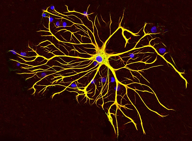
While astrocytes normally help fight β-amyloid plaque deposition, sustained astrocyte reactivity could lead to astrocyte dystrophy, gray matter atrophy, and glucose hypometabolism.

enter image description here

In this new study, scientists Nicholas R Livingston, Paul Edison and their colleagues at Imperial College, London, used the new imidazoline receptor 11C-BU99008 PET tracer to test the hypothesis of a dynamic relationship between the reactivity of astrocytes and neurodegeneration.

They found evidence of increased astrocyte reactivity in patients with pathologically developed amyloid plaques, mainly in the frontal, parietal and occipital regions. These increases were greater in patients with mild cognitive impairment than in patients with Alzheimer's disease.

Further analyzes showed that the lower reactivity of astrocytes in patients with pathologically developed amyloid plaques was associated with both hypometabolism of glucose in the parietal, temporal and frontal lobes and gray matter atrophy in the frontal and temporal lobes.

However, greater deposition of β-amyloid plaques was associated with increased reactivity of astrocytes in primary cortical motor and sensory areas, but decreased reactivity of astrocytes in temporal regions.

11C-BU99008 is a novel PET tracer that binds to I2-BS, the expression of which is associated with the reactivity of astrocytes. There are three main classes of imidazoline receptors: I1 is involved in inhibiting the sympathetic nervous system to lower blood pressure, I2 has functions as yet unclear but is involved in several psychiatric disorders, and I3 regulates insulin secretion. The I2-BS protein located in the brain, is upregulated with healthy aging, and is further increased in Alzheimer's disease.

Different past studies have reinforced the hypothesis that astrocyte reactivity is an early event in the progression of Alzheimer's disease pathology, occurring in response to early amyloid deposition, which usually originates in the frontal lobe. In the early stages, reactive astrocytes have a neuroprotective role, aiding in the clearance of β-amyloid plaques.

In the cohort studied by the scientists, they found weaker binding of 11C-BU99008 in the temporal lobe, which was associated with greater relative progression of amyloid-associated neuropathology, i.e. glucose hypometabolism and gray matter atrophy. Scientists propose that this reduced uptake of 11C-BU99008 in the temporal lobe region reflects astrocyte dystrophy, caused by a chronic pro-inflammatory and neurotoxic amyloid-induced astrocyte phenotype resulting in reduced glycolytic capacity and impairment. secondary neuronal metabolism or cell loss.

There are obvious limits to the study of scientists. First, only a small number of subjects could be examined. A second limitation was the cross-sectional design, which scientists recognize; however, postmortem pathology has the same limitation. The authors' results are therefore best interpreted descriptively and suggesting a hypothetical model, rather than a solid and independent test.

Nonetheless, the consistency of effect directions observed in this study and previous PET 11C-DED studies provide compelling support for the proposed model, where astrocyte reactivity occurs in response to early deposition of β-amyloid plaques, aiding in the clearance of β-amyloid plaques, but where after time the astrocytes become neurotoxic, contributing to reduced tissue activity and cell death associated with cognitive impairment.

Advertisement


This book retraces the main achievements of ALS research over the last 30 years, presents the drugs under clinical trial, as well as ongoing research on future treatments likely to be able stop the disease in a few years and to provide a complete cure in a decade or two.


Please, help us continue to provide valuable information: