Avec le vieillissement les cellules T du système immunitaire adaptatif sont souvent épuisées et/ou deviennent sénescentes. Les personnes atteintes de lymphocytes T dysfonctionnels courent un risque élevé d’infections, de cancer, de maladies chroniques et éventuellement de mortalité.
Un texte récemment publié étudie la relation entre l'inflammation, les altérations du système immunitaire et la maladie d'Alzheimer (maladie d'Alzheimer). Alors que l'état d'esprit commun est de se demander ce qui cause la maladie d'Alzheimer (les bêta-amyloïdes), ce texte adopte une vision plus complexe : le système immunitaire vieillissant étant moins efficace, une deuxième ligne de défense entre en jeu : les bêta-amyloïdes.
Il y a de nombreuses études montrant un lien entre le système immunitaire et la maladie d'Alzheimer. Une inflammation a été observée dans des analyses cérébrales post-mortem de patients atteints de la maladie d'Alzheimer, ainsi que la présence de plaques amyloïdes et d'enchevêtrements neurofibrillaires.
L'utilisation d'anti-inflammatoires non stéroïdiens (AINS) a montré un risque plus faible de démence ou de maladie d'Alzheimer chez les adultes qui les utilisent périodiquement, bien que les résultats des essais cliniques avec les AINS aient été mitigés.
Il y a aussi un lien entre les changements cognitifs et les infections aiguës telles que la septicémie, la méningite et même le COVID-19. De même il y a un lien entre les infections chroniques et le déclin cognitif à long terme. Les herpès virus humains, en particulier le virus de l'herpès simplex-1 (HSV-1) et l'herpèsvirus humain 6 (HHV6), sont considérés comme des contributeurs potentiels à l'inflammation liée à l'infection à l'origine de la maladie d'Alzheimer.
D'autres agents pathogènes comme Porphyromonas gingivalis, Chlamydia pneumoniae et Toxoplasma gondii ont été associés au développement de la maladie d'Alzheimer en raison de leur nature chronique. Les vaccinations contre des maladies comme la grippe, le zona et le BCG ont montré des associations avec une diminution du risque de maladie d'Alzheimer dans diverses populations.
Pour comprendre comment le système immunitaire périphérique est altéré, il est intéressant d'étudier une cohorte vieillissante à différents stades de développement de la maladie d'Alzheimer.
Les scientifiques ont observé des altérations majeures du système immunitaire inné périphérique, dans le sang des membres de la cohorte vieillissante. La cytométrie en flux haute dimension, l'imagerie TEP amyloïde et les tests cognitifs ont été utilisés pour identifier les changements dans les systèmes immunitaires innés et adaptatifs à mesure que la pathologie amyloïde et les symptômes cognitifs se développaient.
Les résultats spécifiques incluent des différences dans les populations de cellules dendritiques, la différenciation des lymphocytes T et la production de cytokines chez les participants amyloïdes positifs, en particulier ceux présentant une déficience cognitive légère.
 Les lymphocytes T matures sont considérés comme immunologiquement naïfs jusqu'à ce qu'ils rencontrent le peptide spécifique dans le contexte d'une molécule d'antigène leucocytaire humain (HLA) que leur récepteur reconnaît. Une fois la reconnaissance de l’antigène effectuée, les cellules reçoivent un signal prolifératif qui conduit à une expansion marquée des lymphocytes T spécifiques de l’antigène et à une réponse inflammatoire. Bien que beaucoup de ces cellules subissent l’apoptose après la réponse initiale, d’autres sont sauvées de la rétraction immunitaire et persistent sous forme de cellules T mémoire. Les lymphocytes T mémoire peuvent répondre rapidement à une nouvelle provocation spécifique d’un antigène et persister dans la circulation à long terme
Lorsque le système immunitaire adaptatif a été examiné, les participants amyloïdes positifs, quel que soit leur état cognitif, présentaient une augmentation de leurs lymphocytes T CD3. Des analyses plus approfondies des lymphocytes T CD4 et CD8 ont révélé que les membres de la cohorte vieillissante présentaient une augmentation du nombre de lymphocytes T de phénotype plus différenciés, par rapport à ceux ayant une cognition normale. C'est à dire qu'à la fois il y avait une moindre production de cellules T naïves et une forte présence de cellules T ayant été en contact avec des pathogènes.
Lorsque la fonction des lymphocytes T a été mesurée, les auteurs ont observé que les lymphocytes T des membres de la cohorte vieillissante avaient augmenté la production de cellules IFN-γ par rapport aux autres participants. L'IFN-γ, ou interféron de type II, est une cytokine essentielle à l'immunité innée et adaptative contre les infections virales, bactériennes et protozoaires. L'IFN-γ est un activateur important des macrophages et un inducteur de l'expression des molécules du complexe majeur d'histocompatibilité de classe II. L’expression aberrante de l’IFN-γ est associée à un certain nombre de maladies auto-inflammatoires et auto-immunes. plusieurs études ont observé une augmentation de l'IFNγ associée à une progression symptomatique plus lente dans la maladie d'Alzheimer.
La protéine de mort cellulaire programmée 1 (PD-1) est une protéine présente à la surface des lymphocytes T et B qui joue un rôle dans la réponse du système immunitaire aux cellules du corps humain en régulant à la baisse le système immunitaire et en favorisant l'autotolérance en supprimant l'activité inflammatoire des lymphocytes T. Cela prévient les maladies auto-immunes, mais cela peut également empêcher le système immunitaire de tuer les cellules cancéreuses. Les auteurs expliquent que les membres de la cohorte vieillissante présentaient une augmentation majeure du nombre de lymphocytes T dépourvus de production de cytokines après la restimulation et exprimaient des niveaux accrus de PD-1 et de Tox, ce qui suggère qu’il s’agit de cellules épuisées.
Compte tenu des nombreux liens entre l'infection, l'inflammation et la maladie d'Alzheimer, ces résultats suggèrent deux modèles dans lesquels les lymphocytes T pourraient être un élément déterminant dans la maladie d'Alzheimer.
Dans le premier modèle, la production d’amyloïde est une réponse aux infections latentes dans la périphérie et le cerveau par les multiples agents pathogènes chroniques que tous les humains sont porteurs. Les individus qui ont une forte fonction des lymphocytes T contrôlent la réplication de ces agents pathogènes et restent cognitivement normaux. Cela expliquerait pourquoi les membres de la cohorte vieillissante, qui possèdent les lymphocytes T les plus fonctionnels, ont toujours niveau cognitif élevé.
- Mais chez les individus qui perdent la fonction des lymphocytes T, les agents pathogènes chroniques se réactivent et surstimulent les réponses innées, en particulier la production d’interféron de type I, conduisant potentiellement à des troubles cognitifs. Les auteurs suggèrent que le rajeunissement des cellules T par des inhibiteurs de points de contrôle immunitaires et d'autres traitements pourrait constituer une thérapie ex vivo plausible pour la maladie d'Alzheimer. En effet, les tests d'inhibiteurs de points de contrôle immunitaires dans le modèle murin 5X FAD de la maladie d'Alzheimer ont donné des résultats prometteurs.
Un modèle alternatif postule que la production de cytokines par les cellules T alors que les participants sont cognitivement normaux entraîne le développement de troubles cognitifs. Cette idée est étayée par une étude récente de Jorfi et ses collègue.
L'étude suggère que le rajeunissement de la fonction des lymphocytes T pourrait constituer un traitement potentiel pour la maladie d'Alzheimer, en particulier les thérapies contre le cancer peuvent suggérer une possibilité. La « neuro-immunothérapie adaptative personnalisée » est une nouvelle méthode permettant de rajeunir et d'améliorer les cellules T de manière sûre et puissante grâce aux neurotransmetteurs et aux neuropeptides, consistant en des protocoles diagnostiques et thérapeutiques personnalisés. Les cellules T rares et/ou dysfonctionnelles du patient sont activées ex vivo une fois par des neurotransmetteurs et/ou neuropeptides présélectionnés, testées et réinoculées au corps du patient.
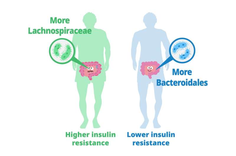 Previous studies have explored the role of gut microbiota in metabolizing nutrients in insulin resistance. This research aims to uncover the mechanisms underlying this relationship using a multi-omics approach. The study analyzes data from 306 individuals without diabetes, focusing on insulin resistance as defined by HOMA-insulin resistance scores.
 Until now, studies on Klotho have primarily focused on its effects on animal models such as mice, rather than directly increasing its levels in primates.
 This condition affects individuals, mostly adults over the age of 50, who physically act out their dreams during sleep, resulting in injuries to themselves or their bed partners. It's also suspected to be involved in premisses of Parkinson's disease. The study, published in the Journal of Neuroscience, presents a novel model that characterizes how REM sleep behavior disorder develops due to neurodegeneration associated with the accumulation of tau protein.
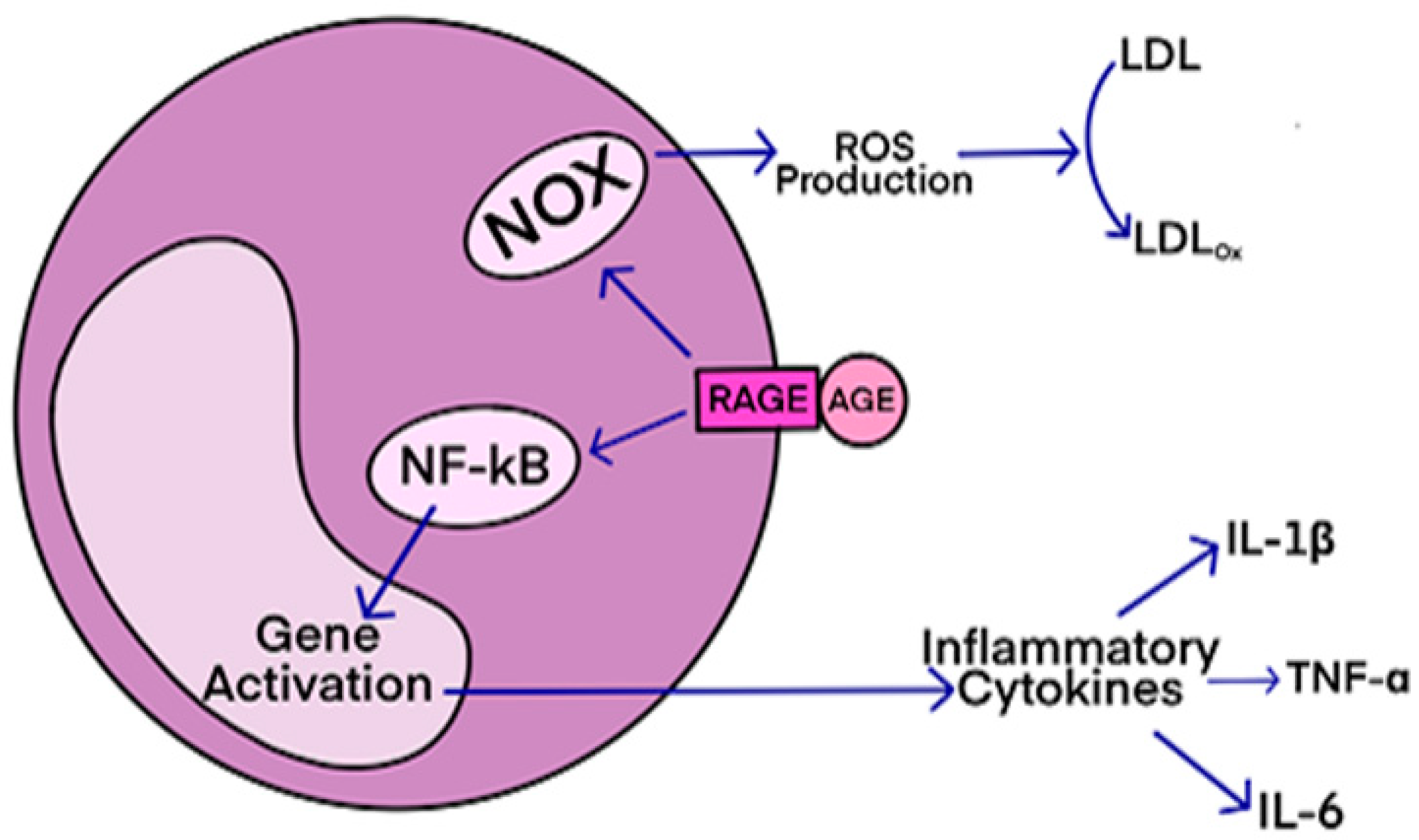 AGEs have diverse structures, but only a limited number have been characterized. Some AGEs are small molecules formed through proteolytic degradation of protein-crosslinked or protein-modified AGEs. Imbalance between the formation and destruction of AGEs, particularly under conditions of oxidative stress, results in excessive accumulation and disease progression.
 Des études précliniques ont montré que pendant le sommeil, l'infiltration du liquide interstitiel et céphalo-rachidien le long des espaces périvasculaires augmente, augmentant ainsi la clairance des solutés interstitiels. Lorsqu'ils sont agrandis, les espaces périvasculaires sont visibles par imagerie par résonance magnétique cérébrale (IRM).
Le volume et le nombre de espaces périvasculaires augmentent avec l'âge.
Ce phénomène est associés à des facteurs de risque vasculaire tels que l'hypertension, des marqueurs de microangiopathie telle que la leucoaraiose et entraine des effets néfastes sur la santé. Une question importante est de savoir si le manque de sommeil est la cause de ces risques vasculaires, ou simplement leur conséquence.
Des études précliniques ont montré que pendant le sommeil, l'infiltration du liquide interstitiel et céphalo-rachidien le long des espaces périvasculaires augmente, augmentant ainsi la clairance des solutés interstitiels. Lorsqu'ils sont agrandis, les espaces périvasculaires sont visibles par imagerie par résonance magnétique cérébrale (IRM).
Le volume et le nombre de espaces périvasculaires augmentent avec l'âge.
Ce phénomène est associés à des facteurs de risque vasculaire tels que l'hypertension, des marqueurs de microangiopathie telle que la leucoaraiose et entraine des effets néfastes sur la santé. Une question importante est de savoir si le manque de sommeil est la cause de ces risques vasculaires, ou simplement leur conséquence.
 Source Wikipedia.
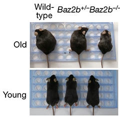 While wild-type mice grew fat with age, animals lacking both copies of the epigenetic reader Baz2b stayed trim, indicating improved mitochondrial function. [Yuan et al., Nature, 2020.]