A study in Norway about drug repurposing

- Posted by admin in English

Although effective symptomatic therapies have been developed for Parkinson's disease (PD), treatments that modify the disease itself still do not exist.

Drug repurposing studies help identify potential disease-modifying treatments. One advantage of drug repurposing is that, since these drugs are already approved, their safety profiles are known. However, further industry investigation is unlikely because companies hesitate to invest in drugs whose intellectual property is owned by others. Generally, they might consider repurposing their own drugs, which extends patent life by claiming new uses. This can also lead to questionable combinations of approved drugs just to file a patent.

In an issue of Neurology, Tuominen et al. publish a nationwide observational cohort study in Norway aiming to identify new candidates for disease modification in Parkinson's disease. They analyzed Norwegian health registries from 2004 to 2020 to identify people with Parkinson's. The study included 14,289 individuals and found 23 drugs among a total of 219 drugs associated with a reduced eight-year mortality risk.

Nonetheless, this reduction remains minor, and there is no proof that these drugs caused improved survival. enter image description here The authors note that, although their findings are exploratory and cannot be directly applied clinically yet, the identified drugs could be considered for future clinical trials. Indeed, funding should be sought for these future trials, and since it is almost always private investors who finance clinical trials, they would require more information before making any commitments.

However, this study has notable strengths compared to similar studies. Tracking specific disease milestones is crucial for evaluating the effects of potential disease-modifying treatments.

For nearly all of the 23 drugs discussed by Tuominen et al., the adjusted eight-year mortality curves for Parkinson's patients and healthy controls diverged and showed different slopes, suggesting these compounds may have a disease-modifying effect. This pattern has not been observed in other trials testing potential disease-modifying drugs.

When interpreting Tuominen et al.'s results, it is important to remember that correlation does not necessarily imply causation. A drug associated with lower mortality does not automatically prove that it caused the reduction.

On the one hand, the decreased mortality might simply reflect that healthier Parkinson's patients are more likely to be prescribed these drugs. For example, patients using tadalafil (for erectile dysfunction) may have better overall health and longer survival.

On the other hand, for the more difficult cases, some physicians may have focused solely on treating essential symptoms rather than prescribing a large number of medications.

Additionally, the number of patients in the "treatment" group is often small; for instance, only 170 patients used Levothyroxine sodium.

Does human pegivirus trigger Parkinson's disease?

- Posted by admin in English

These days, major news stories about neurodegenerative diseases are rare. One headline in the trade press claims: "Harmless Virus Could Trigger Parkinson's Disease."

Most cases of Parkinson's disease are idiopathic, meaning the cause is unknown. However, several genetic mutations can also lead to this neurodegenerative disease, with approximately 20 to 25 percent of cases having a genetic cause. One of these mutations is in the gene encoding LRRK2, which can result in enzyme levels two to three times higher than normal. These mutations are more common in North African, Arab, Berber, Chinese, and Japanese populations. enter image description here

There is some good news coming out on this topic, but I'll talk about another publication.

Every viral infection alters the genetic material of some (but not all) cells in our body. This is how a virus forces the host cell to rapidly produce thousands of copies of itself. In most cases, viruses are first abruptly eliminated by the innate immune system, killing the host cell. The adaptive immune system uses a more intelligent mechanism, producing specific antibodies that bind to the virus and often render it non-infectious. This process is called humoral immunity. The LRRK2 protein is highly expressed in immune system cells, particularly in response to bacterial pathogens like Salmonella.

The hypothesis that some neurodegenerative diseases are caused by viral infections is attractive because it could explain why these diseases appear with age and why they affect the nervous system, as many viruses tend to accumulate there with age.

In a new study, researchers analyzed brains provided by the Rush Alzheimer's Research Center (RADC) in Chicago, from 10 autopsied Parkinson's patients and 14 non-PD patients. They found traces of human pegylated virus (HPgV) in five Parkinson's brains, but none in healthy brains. Human pegylated virus is a virus related to hepatitis C. The virus has also been detected in the cerebrospinal fluid of Parkinson's disease patients, but not in the control group. However, the small sample size makes these results inconclusive.

Next, perhaps seeking stronger evidence, the researchers analyzed blood samples from 1,393 participants in the Parkinson's Progression Markers Initiative, a biological sample bank for Parkinson's disease research. Only about 1% of Parkinson's patients had HPgV in their blood, which is consistent with the infection rate in the general population.

Nevertheless, the scientists say that people infected with the virus exhibited different immune signals, particularly those with a mutation in the LRRK2 gene. They explained that since mutations in the LRRK2 gene are known to influence immune signaling, autophagy, and viral processing, these genotype-specific responses suggest that host genetics and viral interactions could influence immune responses to human pegivirus, promoting neuroinflammation and the development of Parkinson's disease.

At this point, I'm confused; they performed two experiments, one not significant and the other negative, but in the article, the scientists still suggest a possible link between Parkinson's disease and human pegivirus. However, the headline and abstract (which will likely be the only parts colleagues read) are much less definitive: they only suggest that human pegivirus alters the transcriptomic profiles of patients with Parkinson's disease.

This is a far cry from the headlines in the trade press: "Harmless virus might trigger Parkinson's disease."

Perineuronal net modulation in a Parkinson's disease mouse model

- Posted by admin in English

A growing number of studies are exploring the role of perineuronal nets (PNNs) in Parkinson’s disease. PNNs are specialized structures in the brain’s extracellular matrix that limit synaptic plasticity, like a stabilizing "glue" between neurons. While this stabilization helps maintain brain function in adulthood, it also reduces the brain’s ability to rewire or adapt, which is important for learning and recovery after injury. enter image description here 
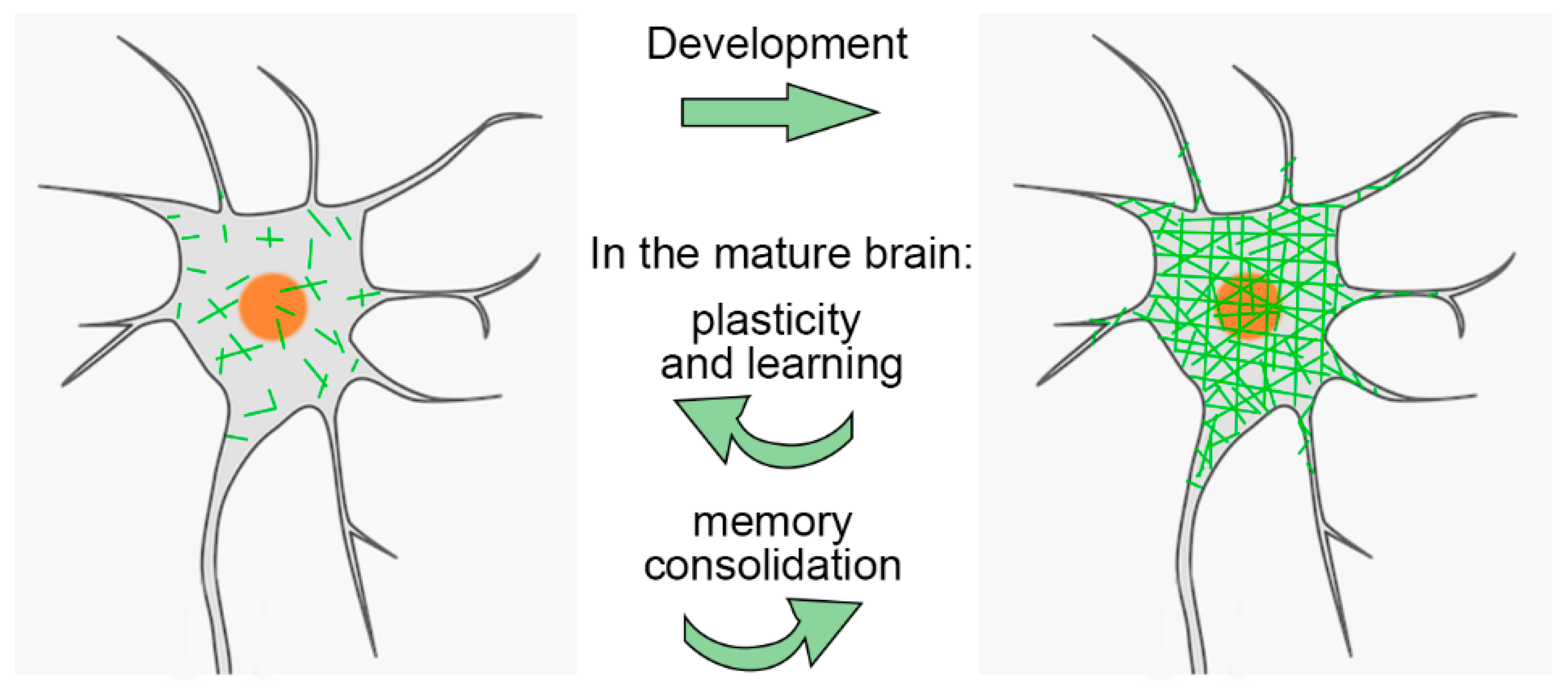
PNNs are especially important during brain development. They help close the "critical period" of heightened plasticity in childhood. Interestingly, while PNNs are degraded in adults, this plasticity can be partially restored. For example, PNN removal can promote recovery in stroke models

PNNs serve multiple functions: protecting neurons from oxidative stress and harmful molecules, regulating plasticity, and helping stabilize long-term memories. Abnormal changes in PNNs have been observed in aging and various neurological conditions, making them a promising target for therapeutic intervention.

In the cerebral cortex, PNNs are primarily found around inhibitory interneurons, particularly those that produce the protein parvalbumin. These interneurons help maintain a balance between excitatory and inhibitory signals in the brain. In Parkinson’s disease, where dopamine-producing neurons in the substantia nigra are lost, this balance is disrupted—suggesting that changes in PNNs may contribute to the disorder.

A recent study investigated the effects of temporarily reducing PNNs in the primary motor cortex (M1) of healthy adult mice using chondroitinase ABC (ChABC). This intervention caused temporary impairments in motor function, suggesting that PNNs contribute to motor stability. Using ChABC makes sense as perineuronal nets are composed of chondroitin sulfate proteoglycans, and ChABC is an enzyme that digests them. Chondroitinase treatment has been shown to allow adults' vision to be restored; moreover, there is some evidence that Chondroitinase could be used for the treatment of spinal injuries.

The researchers created a Parkinson’s disease mouse model by damaging one side of the midbrain of mice with 6-hydroxydopamine (6-OHDA). Two weeks after the lesion, PNN levels dropped in both hemispheres of the motor cortex but returned to normal within five weeks.

The researchers then applied ChABC to reduce PNNs again in the motor cortex and paired this with motor training. This combination improved motor recovery slightly in the Parkinsonian mice. The improvement was linked to an increase in parvalbumin interneurons surrounded by PNNs and a normalization of excitatory signals at their cell bodies. These findings suggest that PNNs respond dynamically—first to the injury and later to therapeutic intervention—and that manipulating them could help restore motor function.

In summary, perineuronal nets in the motor cortex appear to play a subtle but significant role in regulating movement. Modifying their structure could open new avenues for motor rehabilitation in Parkinson’s disease.

Parkinson's disease and acupuncture

- Posted by admin in English

I am somewhat skeptical about the benefits of acupuncture beyond the placebo effect. However, this article suggests it could be beneficial for individuals with Parkinson's disease.
enter image description here This study investigates whether acupuncture has any impact on long-term health outcomes, such as mortality, disease progression, or complications, in individuals newly diagnosed with Parkinson’s disease (PD) in South Korea. Indeed, it is unclear what constitutes an effective acupuncture session for Parkinson's disease, and individuals interested should receive at least one session every two months. It is commonly believed that in PD, reduced mobility due to tremors, postural imbalance, and rigidity likely contributes to poor circulation and decreased gastrointestinal motility, leading to bowel obstructions and impaired swallowing, which can, in turn, result in recurrent aspiration pneumonia. The benefits may arise from the fact that people with PD might find it easier to move.

As is often the case, the Korean authors used a database to select patients and gather information about their health over the following years. They did not see any patients in person; it was purely a matter of processing numbers in the database. The NHIS database contains extensive patient information, including diagnostic codes, healthcare utilization, prescriptions, vital signs, disability grades, sex, age, and socioeconomic factors such as health insurance. However, this database does not provide precise medical details.

Who Was Included in the Study?

The study focused on adults (age 19 or older) newly diagnosed with idiopathic Parkinson’s disease (IPD) between 2012 and 2016.

To ensure a cleaner analysis, the authors excluded individuals diagnosed with Parkinson’s before 2012 or who were disabled at the time of diagnosis. They also excluded patients diagnosed with dementia or who died within a year of their diagnosis. To strengthen the results, they excluded patients who had received acupuncture in the six months preceding their diagnosis.

After applying these exclusions, about 41,000 patients remained. From this group, the researchers employed "propensity score matching" to pair patients from both groups who had similar health and demographic profiles (like age, sex, income, location, and other health conditions). After further exclusions and matching, 6,394 patients remained in each group.

The researchers measured all-cause mortality (death from any cause), tracked for up to six years following diagnosis. They also recorded fractures (such as hip or spine fractures), emergency room visits, and deep brain stimulation (DBS) surgery. These outcomes serve as proxies to assess whether acupuncture is associated with slower disease progression or fewer complications.

How Were Acupuncture and Non-Acupuncture Groups Defined?

Acupuncture was counted only if a person had six or more sessions in the year following diagnosis. This cutoff is based on previous studies suggesting that at least six sessions may be necessary to observe an effect. The acupuncture types included conventional methods as well as specialized forms like ocular or electroacupuncture.

Results:

Overall, the mortality in the acupuncture group (960) was significantly lower than in the control group (1,118). Deaths due to neoplasms (174 vs 234), circulatory (172 vs 208), and digestive diseases were also lower in the acupuncture group. In all cases, the acupuncture group had equal or better results than the control group. However, no significant differences were observed between groups in fracture risk, emergency room visits, or DBS procedures.

Key Takeaways

The study aimed to explore whether acupuncture might influence survival or disease progression in Parkinson’s disease. To achieve this, it utilized a large, high-quality national health dataset and careful matching to compare similar patients. While the text does not cover the results themselves (e.g., whether acupuncture improved survival or reduced complications), the methods ensure that any differences found are likely due to the treatment rather than other health or demographic factors.

How to cope with the deluge of scientific publication?

- Posted by admin in English

At Padiracinnovation, we face a paradox: despite the deluge of scientific publications on ALS, Parkinson's, and Alzheimer's disease, academic and industry scientists seem unable to develop effective drugs.

In fact, the incentive for academic scientists appears to be publishing a large volume of work, as this is nearly their only path to career advancement. Industry scientists publish infrequently, but their work primarily serves to promote their company to potential investors.

How can we differentiate the wheat from the chaff? There are several telltale signs:

  • single author

  • authors who publish more than two papers per year

  • publishing outside of prominent journals, such as in conferences

  • articles based on specific intuition without testing other hypotheses

  • articles concerning a specific drug without explaining the rationale for its initial selection

  • articles making bold claims based on queries to a public database without conducting further research to validate the results and without considering confounding factors

  • articles making outrageous claims, such as "breakthrough in disease X."

In our field, additional indicators exist: a strong publication should report results from human trials, as animal models often prove to be completely ineffective.

Consequently, we find few publications to discuss, even as popular science news organisations report daily on significant advances made toward new drugs.

Abnormal protein aggregation within cells is a recurring phenomenon in Parkinson's disease (PD), Alzheimer's disease (AD), and amyotrophic lateral sclerosis (ALS). Current approaches use antibodies to target these aggregates, but this is a rudimentary approach, as little is known about the causes of their formation, or whether they are the cause or consequence of the disease.

Cells are an incredibly crowded environment, and their molecules undergo Brownian motion, which thwarts their biological function. Making the cell less dense and more soluble would certainly alleviate some molecular problems. There are various approaches, including those that use phase transitions.

Recent research sheds surprising light on the dynamic relationship between mitochondrial activity, ATP levels, and neuronal cytoplasmic fluidity, all of which play a critical role in controlling protein aggregation.

The researchers used mouse giant goblet cell cultures to analyze presynaptic viscosity using real-time confocal microscopy. These cells are characterized by large glutamatergic nerve terminals, ideally suited for real-time imaging. Rather than focusing on individual proteins, the team took a holistic approach, using a technique called fluorescence recovery after photobleaching (FRAP) of soluble green fluorescent protein (cGFP) to assess the overall viscosity of the axonal cytosol.

Cytosolic viscosity can reflect the extent of protein aggregation; Greater aggregation means less free diffusion of cGFP, indicating a more "solidified" cytosol.

Synapses are hotspots for mitochondria, which provide the ATP needed for neurotransmission. By labeling active mitochondria and comparing their location to cGFP mobility, the study revealed that regions with greater mitochondrial activity exhibited higher cytosolic fluidity. This suggests a direct link between ATP production and the maintenance of a more soluble and functional presynaptic environment.

To further investigate this, the team inhibited mitochondrial function using FCCP and other mitochondrial blockers. As ATP production decreased, cGFP diffusion decreased sharply, suggesting that the cytosol was becoming more viscous due to protein aggregation. It is important to note that this effect was specific to mitochondrial inhibition: blocking glycolysis had little effect.

Even components of the synaptic release mechanism, such as synaptic vesicles (SVs) and active zones (AZs), exhibited reduced mobility under mitochondrial stress, reinforcing the idea that energy depletion disrupts the fluid phase of the cytoplasm.

To test whether ATP could restore the altered cytosol state, the researchers administered ATP directly to neurons. They found that ATP not only restored cGFP diffusion but also reduced the size and number of protein aggregates. To test whether enhancing endogenous ATP production could mitigate the protein aggregation linked to mitochondrial dysfunction, the researchers turned to NMN, a molecule known to boost NAD⁺ levels and support mitochondrial health.

They treated neurons with NMN and observed the following key outcomes:

Partial restoration of cytoplasmic fluidity: In neurons with compromised mitochondrial activity (such as those derived from PARK2 or TDP-43 mutant patients), NMN treatment significantly improved the diffusion of soluble proteins like cGFP. While not as dramatic as direct ATP infusion, NMN nonetheless reduced cytosolic viscosity.

Reduction in aggregate burden: In both mouse neurons under mitochondrial stress and hiPSC-derived human neurons from neurodegenerative disease patients, NMN treatment lowered the accumulation of insoluble protein aggregates.

Improved ATP levels: NMN supplementation helped increase intracellular ATP concentrations, presumably by enhancing mitochondrial NAD⁺-dependent enzymatic activity, which supports oxidative phosphorylation.

These results suggest that NMN supports the same protective pathway as ATP, but indirectly, by restoring mitochondrial capacity to generate ATP and maintain a more fluid intracellular environment.

The mechanism appears to be biophysical rather than biochemical: ATP acts as a hydrotrope, a molecule that keeps other proteins dissolved and prevents them from forming aggregates.

The researchers then examined whether this principle held true for specific proteins involved in neurodegenerative diseases, including:

  • α-synuclein (mutant SNCA and SNCA-A53T), PARK2 – Parkinson's disease

  • APP, Amyloid, Tau – Alzheimer's disease

  • TDP-43 – ALS

These purified proteins were able to undergo liquid-liquid phase separation (LPS) and form condensates in vitro. ATP was able to dissolve many of these condensates in a concentration-dependent manner, although mutant or misfolded versions (e.g., SNCA-A53T) required higher ATP concentrations to dissolve.

When the aggregates were left to incubate for longer, some (notably SNCA-A53T) began to form protofibrils, elongated, fibril-like structures similar to those observed in real-life pathology. Here again, ATP could reverse this phenomenon, but with reduced efficiency.

Even under crowded conditions (mirrored by the addition of PEG), ATP retained some ability to prevent or dissolve aggregates, although the effect was less potent.

The team then studied neurons derived from Human induced pluripotent stem cells (hiPSCs) from patients with Parkinson's disease (PARK2 mutation) and ALS (TDP-43 mutation). These neurons exhibited reduced cytosolic fluidity, lower ATP levels, and greater protein aggregation than healthy controls.

This supports the idea that ATP deficiency and mitochondrial dysfunction contribute to the condensation of pathogenic proteins in human neurodegenerative diseases.

Implications for Drug Development

This research redefines our approach to therapeutic targets in neurodegenerative diseases. Instead of seeking to eliminate aggregates after their formation, we could:

  • Target mitochondrial function to preserve ATP production at synapses.

  • Use small molecules that mimic the hydrotropic effects of ATP to maintain cytoplasmic fluidity.

  • Develop drugs that prevent the formation of LPS (lipoproteinases) of key disease proteins by improving their solubility.

ATP itself is not a drug molecule in the traditional sense, but these results open new avenues for small molecules capable of acting like ATP to maintain protein solubility or prevent aggregate formation at an early stage.

Conclusion

Neurodegenerative diseases are often viewed from a genetic or protein perspective, but this study provides a biophysical perspective: the physical state of the cytosol itself is crucial. If cells cannot maintain a fluid and soluble environment, primarily due to energy deficiency, aggregation may become inevitable.

This is not just about treating symptoms or even eliminating aggregates afterward. It is about preserving the cellular environment so that neurons can withstand stress and maintain their function. As the field continues to explore how biophysical properties such as viscosity, solubility, and phase separation interact with disease, the role of ATP may prove central, not only as a fuel, but also as a key regulator of neuronal health.

Differences in disease treatment between countries are evidence that medicine is not an exact science (if there ever was one). For example, it has been shown in certain cancers that crossing a state border can offer a better chance of survival. The article that is the subject of this post takes us to China in Taizhou, in the Zhejiang province.

It seems that in Asia (China, Japan) we talk about Parkinson's disease with dementia, as distinct from Lewy body disease. This dementia is managed with donepezil, which is not done in the West where this drug is rather used for Alzheimer's disease.

Donepezil is one of those drugs with unpleasant side effects that sometimes lead to patients abandoning them.

Murine NGF (nerve growth factor) has been licensed in China since 2003. It appears to improve patient outcomes for several nervous system diseases. This is important because few drugs can treat nervous system diseases. Unfortunately, research and clinical use outside of China are limited.

Doctors in Taizhou wanted to investigate the clinical efficacy of donepezil combined with nerve growth factor (NGF) in the treatment of Parkinson's disease (PD) dementia and its potential impact on serum adiponectin (APN) and soluble tumor necrosis factor receptor-1 (sTNFR-1) levels.

Clinical data from 140 PD patients treated at Taizhou People's Hospital from March 2021 to December 2023 were retrospectively analyzed. Patients were grouped according to the treatment received. Patients receiving donepezil alone (n = 68) were in the Donepezil group, and patients treated with a combination of donepezil and NGF (n = 72) were assigned to the Donepezil and NGF group.

The overall efficacy of the combination therapy was superior to that of donepezil alone treatment. enter image description here The authors focused on adiponectin, an adipocytokine, i.e. a molecule produced by adipose tissue, which is involved, among other things, in the regulation of lipid and glucose metabolism. Adiponectin modulates inflammatory cascades by modifying the action and production of inflammatory cytokines, but the link between adiponectin and Parkinson's disease is not obvious unless we consider that Parkinson's disease is due to a metabolic disorder. The relationship with the soluble tumor necrosis factor receptor (sTNFR) is even less obvious. Nothing in the article explains why these two molecules were studied.

The serum APN levels after treatment in the donepezil and NGF group were significantly higher than in the donepezil alone group, while the sTNFR-1 level was significantly lower. There was no significant difference in the incidence of adverse events between the two groups.

In conclusion, the combined treatment regimen of donepezil and NGF is more effective than donepezil monotherapy in improving cognitive function, neurological function, and severity of the condition in patients with Parkinson's disease with dementia, and is associated with suppression of the inflammatory response without a significant increase in the incidence of adverse events. Hopefully, these studies will be considered in the Western world.

This is a short post about this publication with an interesting title.

I thought it was about the acute anxiety of carers when their loved one could not eat anymore, actually, it is about how doctors in British hospitals manage patients who could not take their medication orally.

The alternatives are either a soluble medication, a topical patch, or a nasogastric tube.

I wonder if there is not a more urgent problem for patients that could not swallow, than taking their medication.

I am pleased that two new articles bear a similar message: That neurodegenerative diseases can't be understood with the paradigm acquired with communicable diseases.

This paradigm tells us that as the pathogen is quite homogeneous, so is the disease phenotype. This is indeed wrong for non-communicable diseases like cancers and is even wrong for some communicable diseases like COVID-19 where the pathogens have heavily mutated.

One of these articles is "Serena Verdi et al, Personalizing progressive changes to brain structure in Alzheimer's disease using normative modeling".

The authors looked at 3233 brain scans. The number of non-standard brain structures increased over time in people with Alzheimer's disease. Patterns of change in outliers varied markedly between individual patients with Alzheimer's disease.

The authors say: "I think we need to pivot towards a new way of thinking to get away from the idea that this (brain) area is important, this area isn't". The big picture and the individual variability contained within it, is what counts.". Some of this individual variability may stem from the fact that many people with Alzheimer's have more than one cause of cognitive illness.

This idea that "we need to pivot towards a new way of thinking to get away from the idea that this (brain) area is important, this area isn't" is certainly important when we think of other neurodegenerative diseases such as ALS or Parkinson's disease.

Our knowledge about Parkinson's disease is limited. The official narrative is that Parkinson's disease is characterized by progressively expanding nerve cell death originating in substantia nigra, a midbrain region that supplies dopamine to the basal ganglia, a system involved in voluntary motor control. The cause of this cell death is poorly understood but involves alpha-synuclein aggregation into Lewy bodies within the neurons. Substantia nigra is really a tiny part of the brain, and other studies have already revealed a wider involvement in the brain. I guess we would make significant progresses in the disease knowledge if we acknowledge that if alpha-synuclein is involved in Parkinson's disease, it is unlikely its effects are limited to a tiny portion of the brain, as alpha-synuclein is abundant in the brain, while smaller amounts are found in the heart, muscle and other tissues and Lewy bodies are in the midbrain and the cortex.

Another interesting article is: "Ophthalmate is a new regulator of motor functions via CaSR: implications for movement disorders".

While the official narrative is that Parkinson's disease is characterized by neuronal death in substantia nigra, which supplies dopamine to the basal ganglia, a system involved in voluntary motor control, it has been known for decades that robust motor activity can happen in Parkinson's mouse models when L-DOPA conversion to dopamine is blocked! The motor improvement is larger than in the conventional case where L-DOPA is metabolized in dopamine. The authors hypothesized that there was an alternative pathway or mechanism, independent of dopamine signaling.

The authors sought to determine the metabolites associated with the pronounced hyperactivity observed. They observed that the peak in motor activity induced by inhibiting L-DOPA conversion into dopamine in Parkinson’s disease mice was associated with a surge (20-fold) in brain levels of the tripeptide ophthalmic acid (also known as ophthalmate in its anionic form). When they administered ophthalmate directly into mice's brains, it rescued motor deficit in a dose-dependent manner.

The team investigated the molecular mechanisms underlying ophthalmate’s action and discovered, that ophthalmate binds to and activates the calcium-sensing receptor (CaSR). To strengthen their findings, they verified that a CaSR antagonist inhibits the motor-enhancing effects of ophthalmate.

A link between Parkinson's disease and the calcium-Sensing Receptor is interesting as CaSR also Mediates β-Amyloid production. There is only one other publication that explicitly links the disease to CaSR. Calcium acts in a number of signal transduction pathways as second messengers, so maybe it is not wise to read too much about a link between Parkinson's disease and CaSR, but finding that lack of dopamine is not the main cause of Parkinson's disease, is a major finding.

Uric Acid in Parkinson Disease: What is the Connection?

- Posted by admin in English

As loving standards progressed during the last century, the diet became richer, and health conditions, such as gout, which were usually associated with people with high living standards became increasingly more common. It was soon recognized that acid uric imbalance was associated, not only with gout, but with a range of diseases. Most striking associations are inverse correlations as they should teach us something. One such inverse correlation is between elevated acid uric levels and Parkinson's disease. On the contrary, there is an association between low levels of acid uric and patients with Parkinson's disease, or multiple sclerosis. Yet something related to Parkinson's disease may cause low uric acid (reverse causation). enter image description here Indeed, Parkinson’s disease is characterized by the loss of dopaminergic neurons in the substantia nigra pars compacta and the presence of Lewy bodies and Lewy neurites, which mainly consist of aggregates of α-synuclein. It is believed that α-synuclein aggregates poison the brain's cells and indeed especially this tiny part of the brain named "substantia nigra". Yet like other protein aggregates, they may form to protect the brain against some external aggression or stressing event. So the biological mechanisms underlying α-synuclein relationships with dopaminergic neurons have never been firmly established.

While the most frequently proposed mechanism for uric acid's inverse association with Parkinson's disease is that it is an antioxidant, however, clinical trials aiming to raise uric acid levels failed to slow the progression of Parkinson's disease. Then some scientists argued that many studies have pointed to mitochondrial dysfunction in Parkinson's disease. One of the major functions of mitochondria is producing energy in the form of ATP, which is quantitatively the most abundant of all purines in the body. As serum uric acid is a by-product of purine metabolism, mitochondrial dysfunction, and energy failure in Parkinson's disease may lead to low serum uric acid levels. An excellent review can be found here.

There is ample evidence that neurons can internalize extracellular aggregates by endocytosis. Some studies have shown that acid uric inhibits α-syn endocytosis by neurons thereby limiting the progression of the disease.

To precise the relation between Parkinson's disease and uric acid, a study shows that factors other than the purine metabolic system might influence CSF values of uric acid and that purine recycling pathways may be impaired. enter link description here

The current study found a significant reduction in hypoxanthine and inosine levels in the CSF of patients with PD but not in the serum. This small study, published in Nature Parkinson's Disease Journal, was published in preprints last year. Basically, it confirms previous findings such as those reported in the 2023 review above.


Please, help us continue to provide valuable information: