Abnormal protein aggregation within cells is a recurring phenomenon in Parkinson's disease (PD), Alzheimer's disease (AD), and amyotrophic lateral sclerosis (ALS). Current approaches use antibodies to target these aggregates, but this is a rudimentary approach, as little is known about the causes of their formation, or whether they are the cause or consequence of the disease.

Cells are an incredibly crowded environment, and their molecules undergo Brownian motion, which thwarts their biological function. Making the cell less dense and more soluble would certainly alleviate some molecular problems. There are various approaches, including those that use phase transitions.

Recent research sheds surprising light on the dynamic relationship between mitochondrial activity, ATP levels, and neuronal cytoplasmic fluidity, all of which play a critical role in controlling protein aggregation.

The researchers used mouse giant goblet cell cultures to analyze presynaptic viscosity using real-time confocal microscopy. These cells are characterized by large glutamatergic nerve terminals, ideally suited for real-time imaging. Rather than focusing on individual proteins, the team took a holistic approach, using a technique called fluorescence recovery after photobleaching (FRAP) of soluble green fluorescent protein (cGFP) to assess the overall viscosity of the axonal cytosol.

Cytosolic viscosity can reflect the extent of protein aggregation; Greater aggregation means less free diffusion of cGFP, indicating a more "solidified" cytosol.

Synapses are hotspots for mitochondria, which provide the ATP needed for neurotransmission. By labeling active mitochondria and comparing their location to cGFP mobility, the study revealed that regions with greater mitochondrial activity exhibited higher cytosolic fluidity. This suggests a direct link between ATP production and the maintenance of a more soluble and functional presynaptic environment.

To further investigate this, the team inhibited mitochondrial function using FCCP and other mitochondrial blockers. As ATP production decreased, cGFP diffusion decreased sharply, suggesting that the cytosol was becoming more viscous due to protein aggregation. It is important to note that this effect was specific to mitochondrial inhibition: blocking glycolysis had little effect.

Even components of the synaptic release mechanism, such as synaptic vesicles (SVs) and active zones (AZs), exhibited reduced mobility under mitochondrial stress, reinforcing the idea that energy depletion disrupts the fluid phase of the cytoplasm.

To test whether ATP could restore the altered cytosol state, the researchers administered ATP directly to neurons. They found that ATP not only restored cGFP diffusion but also reduced the size and number of protein aggregates. To test whether enhancing endogenous ATP production could mitigate the protein aggregation linked to mitochondrial dysfunction, the researchers turned to NMN, a molecule known to boost NAD⁺ levels and support mitochondrial health.

They treated neurons with NMN and observed the following key outcomes:

Partial restoration of cytoplasmic fluidity: In neurons with compromised mitochondrial activity (such as those derived from PARK2 or TDP-43 mutant patients), NMN treatment significantly improved the diffusion of soluble proteins like cGFP. While not as dramatic as direct ATP infusion, NMN nonetheless reduced cytosolic viscosity.

Reduction in aggregate burden: In both mouse neurons under mitochondrial stress and hiPSC-derived human neurons from neurodegenerative disease patients, NMN treatment lowered the accumulation of insoluble protein aggregates.

Improved ATP levels: NMN supplementation helped increase intracellular ATP concentrations, presumably by enhancing mitochondrial NAD⁺-dependent enzymatic activity, which supports oxidative phosphorylation.

These results suggest that NMN supports the same protective pathway as ATP, but indirectly, by restoring mitochondrial capacity to generate ATP and maintain a more fluid intracellular environment.

The mechanism appears to be biophysical rather than biochemical: ATP acts as a hydrotrope, a molecule that keeps other proteins dissolved and prevents them from forming aggregates.

The researchers then examined whether this principle held true for specific proteins involved in neurodegenerative diseases, including:

  • α-synuclein (mutant SNCA and SNCA-A53T), PARK2 – Parkinson's disease

  • APP, Amyloid, Tau – Alzheimer's disease

  • TDP-43 – ALS

These purified proteins were able to undergo liquid-liquid phase separation (LPS) and form condensates in vitro. ATP was able to dissolve many of these condensates in a concentration-dependent manner, although mutant or misfolded versions (e.g., SNCA-A53T) required higher ATP concentrations to dissolve.

When the aggregates were left to incubate for longer, some (notably SNCA-A53T) began to form protofibrils, elongated, fibril-like structures similar to those observed in real-life pathology. Here again, ATP could reverse this phenomenon, but with reduced efficiency.

Even under crowded conditions (mirrored by the addition of PEG), ATP retained some ability to prevent or dissolve aggregates, although the effect was less potent.

The team then studied neurons derived from Human induced pluripotent stem cells (hiPSCs) from patients with Parkinson's disease (PARK2 mutation) and ALS (TDP-43 mutation). These neurons exhibited reduced cytosolic fluidity, lower ATP levels, and greater protein aggregation than healthy controls.

This supports the idea that ATP deficiency and mitochondrial dysfunction contribute to the condensation of pathogenic proteins in human neurodegenerative diseases.

Implications for Drug Development

This research redefines our approach to therapeutic targets in neurodegenerative diseases. Instead of seeking to eliminate aggregates after their formation, we could:

  • Target mitochondrial function to preserve ATP production at synapses.

  • Use small molecules that mimic the hydrotropic effects of ATP to maintain cytoplasmic fluidity.

  • Develop drugs that prevent the formation of LPS (lipoproteinases) of key disease proteins by improving their solubility.

ATP itself is not a drug molecule in the traditional sense, but these results open new avenues for small molecules capable of acting like ATP to maintain protein solubility or prevent aggregate formation at an early stage.

Conclusion

Neurodegenerative diseases are often viewed from a genetic or protein perspective, but this study provides a biophysical perspective: the physical state of the cytosol itself is crucial. If cells cannot maintain a fluid and soluble environment, primarily due to energy deficiency, aggregation may become inevitable.

This is not just about treating symptoms or even eliminating aggregates afterward. It is about preserving the cellular environment so that neurons can withstand stress and maintain their function. As the field continues to explore how biophysical properties such as viscosity, solubility, and phase separation interact with disease, the role of ATP may prove central, not only as a fuel, but also as a key regulator of neuronal health.

It's a bit sad that scientists studying FTD think of themself as "dementia scientists" while scientists studying ALS or Parkinson's disease think they belong to a motor disorders category and motor neurons specialists for ALS scientists, while many neurodegenerative diseases share a lot of molecular and physiologic characteristics. At least these cases are often those of aged people and they involve mislocated and misfolded protein aggregates.

So many scientists from the Memory and Aging Center at the University of California, were motivated to study the impact of age on neurodegenerative diseases:

  • They tell that age is the biggest risk factor for dementia, which is a way to present aging as a cause of neurodegenerative diseases, not simply a comorbidity.
  • Most dementia cases (>75%) involve multiple types of brain pathologies, which implies again that those pathologies are not diseases in the same sense as communicable diseases where usually there is a single pathogen and removing this pathogen more or less (not always) restore health.
  • Previous animal experiments showed that exchanging blood between young and old animals could affect brain aging (called "heterochronic blood experiments"). This is a controversial topic as some ultra-rich people already buy young blood of unclear origin. Identifying the detrimental substances and those that are beneficial would help human society as a whole.
  • While individual blood factors had been identified in animal studies, their relevance to human disease wasn't well understood

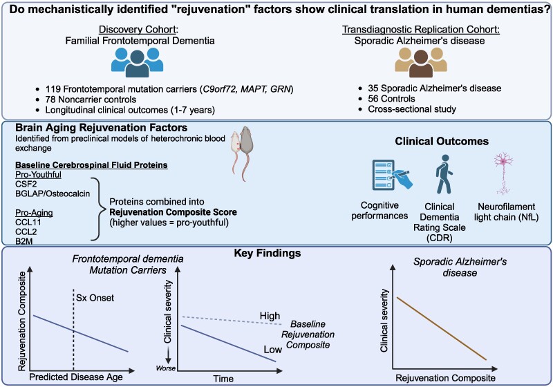
This study involved the direct examination of persons in two cohorts: A longitudinal study of people with genetic frontotemporal dementia (FTD) and healthy controls. A cross-sectional study of people with sporadic Alzheimer's disease and controls.

  • Discovery Cohort (ALLFTD Study): 119 people with FTD genetic mutations (37 MAPT, 33 GRN, 49 C9orf72) 78 healthy controls without mutations This was a longitudinal study (participants were followed over time) Participants had on average 3 annual evaluations (ranging from 1-7 visits) About half (52) of the mutation carriers were asymptomatic at the beginning of the study.

  • Replication Cohort (Stanford ADRC): 35 people with Alzheimer's disease 56 clinically normal older adults This was a cross-sectional study (participants were NOT followed over time)

For both groups, the scientists collected: - Cerebrospinal fluid (CSF) through lumbar punctures - Comprehensive cognitive tests - Functional assessments (rated by caregivers) - Blood or CSF samples for NfL (a marker of neurodegeneration)

The scientists identified five specific proteins from previous animal studies: - Could cross the blood-brain barrier - Were measurable in human samples - Had shown effects on brain aging

These proteins included: Three "pro-aging" factors: CCL11, CCL2, B2M Two "pro-youthful" factors: CSF2 and BGLAP

They found that people with FTD mutations had lower levels of "rejuvenation proteins". Higher levels of these proteins were associated with slower disease progression The protective effect was seen across multiple cognitive domains. The effect was similar regardless of which specific FTD mutation people had

Similar protective associations were found in Alzheimer's disease. Higher levels of these proteins were associated with better cognitive performance and functional status. The effect was particularly strong for memory performance.

  • CCL11 is a small cytokine belonging to the CC chemokine family. CCL11 selectively recruits eosinophils by inducing their chemotaxis, and therefore, is implicated in allergic responses. Increased CCL11 levels in blood plasma are associated with aging. Exposing young mice to CCL11 or the blood plasma of older mice decreases their neurogenesis and cognitive performance on behavioral tasks.
  • CCL2, another cytokine, is implicated in pathogeneses of several diseases characterized by monocytes (a type of leukocyte or white blood cell) infiltrates, such as psoriasis, rheumatoid arthritis, and atherosclerosis
  • B2M is a component of MHC class I molecules. MHC class I function is to display peptide fragments of proteins from within the cell to cytotoxic T cells. Systemic B2M accumulating in aging blood promotes age-related cognitive dysfunction and impaired neurogenesis. In addition, it promotes beta-amyloid aggregation and neurotoxicity in models of Alzheimer’s disease.
  • CSF2 is a monomeric glycoprotein secreted by macrophages, T cells, mast cells, natural killer cells, endothelial cells, and fibroblasts, that functions as a cytokine.
  • Osteocalcin, also known as (BGLAP), is a protein hormone found in bone. Numerous recent studies have revealed bidirectional crosstalk between the brain (and Alzheimer's disease) and bone health.

The publication does not mechanistically explain these "pro-aging" and "pro-youthful" factors. It may suggest that it pays to have a low inflammation level and to be physically active. enter image description here The results appear relatively reliable because the scientists found similar effects in two different types of dementia. The effects were seen across multiple measures (cognitive, functional, and biological markers).
This research could impact drug development in several ways: - It suggests targeting multiple proteins simultaneously might be more effective than single-target approaches - It identifies specific proteins that could be therapeutic targets - It demonstrates these effects in humans, making it more likely to translate into effective treatments - The proteins are measurable in the blood, which could make treatment monitoring easier and safer than the very intrusive CSF sampling.

While not directly studied, this research could be relevant to ALS because ALS shares some biological mechanisms with FTD (they're often considered part of the same disease spectrum).

The research suggests a new paradigm for treating neurodegenerative diseases by targeting multiple age-related factors simultaneously, rather than focusing on single disease-specific pathologies. This could be particularly relevant for diseases like ALS where multiple mechanisms contribute to disease progression.

Short news in neurodegenerative research

- Posted by admin in English

We decided to use a new post format in 2025. It would propose aggregated short news instead of dedicating a post per publication.

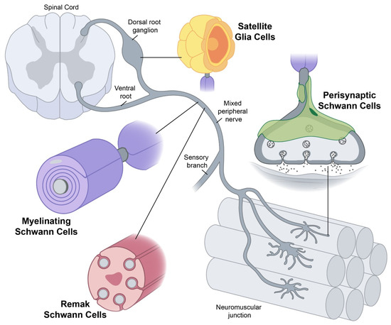
Scientists who publish on neurodegenerative diseases often ignore the fact that neuron-type cells comprise only half of cells in the central nervous system. So it's fresh air to read a review on Schwann cells involvement in ALS. enter image description here Yet conceptually associating Schwann cells and ALS is not common, ALS is a disease of the central nervous system (upper motor neurons in the brain and spine) while Schwann cells are located in the peripheral nervous system (lower motor neurons with their bodies from the spine and terminating in muscles). This does not mean there are no relations between the two types of cells, and it's a common view now that neurons are not independent, self-sufficient entities and that they are cared for by a large number of other cell types.
In a similar vein, Uruguayan scientists were interested in astrocytes' health. Astrocytes are the main kind of motor neuron supportive cells, if they ill behave, motor neurons die and it happens that they can switch between several behaviors. The scientists thought that metabolic reprogramming could occur in astrocytes following damage, and it significantly influences the progression of ALS pathology. Metabolic reprogramming, which involves changes in mitochondrial activity, within glial cells may provide valuable insights for developing innovative therapeutic approaches to mitigate neuronal damage.

It's no new but another study finds common molecular features between ALS and Parkinson's diseases. This reinforces the idea that sporadic neurodegenerative diseases are not clearly delineated diseases as in medical books. On the contrary, these medical classifications just describe symptoms belonging to a spectrum shared by many sporadic neurodegenerative diseases and aging.

Scientists in Taiwan studied the effects of isofraxidin on motor performance changes in chemically induced (lipopolysaccharide) Parkinson's disease in mice.

Isofraxidin is isolated from Eleutherococcus senticosus. Eleutherococcus senticosus, as many berries, is itself loaded with chemical components and provokes adverse effects in some people. What makes them study this plant is not disclosed. Still, as often it's probably because it is used in traditional medicine, and there were some interesting scientific studies on its effects on neurological disease. Isofraxidin pre-treatment significantly improved lipopolysaccharide-induced motor dysfunction, as evidenced by better performance in the rotarod, pole-climbing, and beam-walking tests. Does this prove anything? I am not sure, there are many articles that isofraxidin protects against lipopolysaccharide-induced diseases, the scientists most probably knew that when they planned their experiment.

This post is about an interesting hypothesis. Hypotheses abound, yet few a convincing.

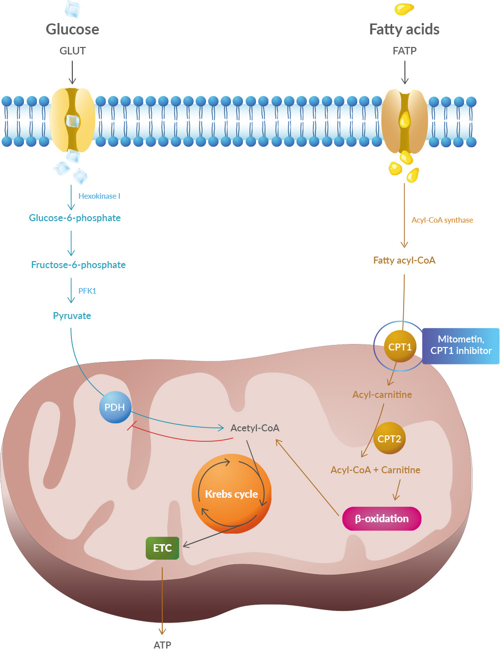
Half of patients with Alzheimer's disease, Parkinson's disease, or ALS have insulin resistance. Obesity and diabetes have been linked to neurodegenerative diseases like multiple sclerosis (MS), Alzheimer's (AD), and Parkinson's (PD). This means the cells of their body cannot let the glucose enter them. Glucose is the main energy source as it is converted into ATP. Glucose is for short-term (day) energy needs. Another source of energy is lipids (fat). Lipids are even more dense than glucose energy-wise.

The body needs an enormous amount of energy. With all the lipids in the body of a healthy person, you could charge two Tesla cars! The brain (a part of the CNS) needs 20% of all energy intake.

A new paper argues that cells shift their metabolism from glucose to lipids under stressors. It tells that one notable distinction between glucose and lipid metabolism is in the quantity of oxygen required to generate each ATP molecule. Lipid metabolism needs two times more oxygen than glucose metabolism. The result is two times more damaging ROS (a by-product of metabolism). enter image description here Studies have shown that oxidative stress and endoplasmic reticulum stress are correlated and can lead to protein misfolding (Abramov et al., 2020). Accumulation of misfolded proteins causes cellular damage and mitochondrial dysfunction and is associated with a range of neurodegenerative diseases, including ALS (misfolded SOD1, TDP-43, C9orf72) (McAlary et al., 2020), Parkinson's disease (misfolded α-synuclein) and Alzheimer disease (misfolded Aβ and Tau) (Abramov et al., 2020).
It explains also the accumulation of iron in patients' brains: To transport oxygen the blood cells need iron, and as the glucose in the blood is not absorbed in cells, it induces a change in microbiota.

It's also well known that SCFAs (including butyrate) have a positive effect on neurodegenerative diseases by their action on microbiota. SCFAs help to restore glucose as the preferred energy substrate. Authors say there are other means to restore glucose as the main source of energy.

What to think about this paper? First, some authors belong to a biotech so we can expect they want to promote their drug: Mitometin. Second, this is a review, this is not even a pre-clinical study, yet some of the authors were involved in pre-clinical studies on this topic. Other groups have written on this topic. What to make of this? Acetyl-CoA carboxylase might be of interest as they produce malonyl-CoA which inhibits the CPT1 gene that regulates lipid metabolism. B7 vitamin is known to convert acetyl-CoA to malonyl-CoA for fatty acid synthesis.

RAGE Inhibitors in Neurodegenerative Diseases

- Posted by admin in English

This post is about a review of the contribution of Advanced Glycation Endproducts to Neurodegenerative Diseases.

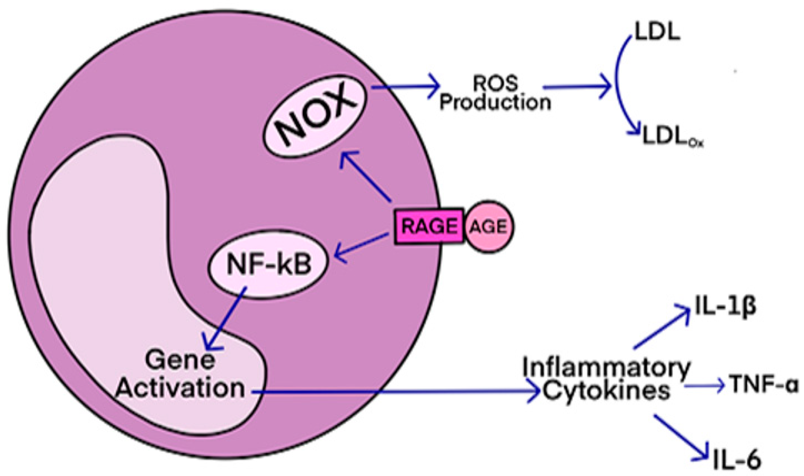
Advanced Glycation Endproducts (AGEs) are formed through non-enzymatic reactions between proteins, aminoglycosides, amino-terminal lipids, and reducing sugars like D-glucose. This process involves Amadori rearrangements and oxidative modifications. The accumulation of AGEs, especially under conditions of elevated oxidative stress, leads to various diseases. enter image description here AGEs have diverse structures, but only a limited number have been characterized. Some AGEs are small molecules formed through proteolytic degradation of protein-crosslinked or protein-modified AGEs. Imbalance between the formation and destruction of AGEs, particularly under conditions of oxidative stress, results in excessive accumulation and disease progression.

Some of the well-characterized AGEs include pentosidine, glucosepane, Argpyrimidine, and Nε-(carboxymethyl)lysine (CML). The imbalance between the formation and destruction of AGEs, triggers a cascade of signaling events, inflammation, and oxidative stress. This inflammatory signaling cascade is associated with various neurological diseases, including Alzheimer’s disease (AD), amyotrophic lateral sclerosis (ALS), diabetic neuropathy, diabetes, and atherosclerosis.

Other endogenous ligands are also involved, such as high mobility group box1 (MGB1) proteins. Additionally, exogenously ingested AGEs contribute to disease onset. The formation and accumulation of AGEs overwhelm the body's detoxification mechanisms under conditions of enhanced oxidative stress, exacerbating neurodegenerative diseases and other inflammatory-associated conditions.

RAGE (receptor for advanced glycation end-products) is a receptor that interacts with AGE (advanced glycation end-products)-derived ligands, such as CML, CEL, and MG-H1. The ligands bind to specific residues on the receptor, initiating signal transduction and pro-inflammatory signaling events.

RAGE antagonists, either endogenous or exogenous compounds, bind to RAGE and attenuate the binding interactions between AGE and RAGE, thereby preventing disease progression. These antagonists have shown potential for treating neurodegenerative diseases, diabetes, atherosclerosis, and cancers. Some small molecule-based RAGE inhibitors, like FPS-ZM1 and Azeliragon, have entered clinical trials, but none have been FDA-approved yet.

FPS-ZM1 has shown selective binding to RAGE and inhibits the formation of Aβ peptides, which are associated with brain damage in diseases like Alzheimer's. RAGE inhibitors also have potential therapeutic applications in diabetic nephropathy, cancer cell metastasis, and Parkinson's disease.

Azeliragon, currently in phase 3 clinical trials, has demonstrated decreased levels of Aβ plaques in the brain, reduces inflammation, and slower cognitive decline in Alzheimer's patients. Other compounds, such as urolithin and its analogs, have shown comparable RAGE inhibition to Azeliragon.

RAGE antagonists are also considered as therapeutics for diabetic neuropathy and retinopathy. Inhibitors of the cytoplasmic tail of RAGE (ct-RAGE) have been investigated and shown to block AGE/RAGE-mediated signaling events effectively. Some of these small molecule antagonists have structural similarities to FPS-ZM1 and have demonstrated potential for treating neurological disorders and diabetic complications.

Dietary AGEs, especially from animal-derived foods cooked at high temperatures, can also contribute to AGE accumulation and the development of diseases. RAGE antagonists, AGE inhibitors, and soluble RAGE (sRAGE) have shown promise in the treatment of age-related pathologies. Polyphenolic compounds can attenuate AGE formation and its toxic effects by reducing oxidative stress.

Therapeutic interventions targeting the AGE-RAGE axis may provide effective treatment for neurodegenerative diseases, diabetes, atherosclerosis, and cancers.

In summary, RAGE antagonists show promise as therapeutics for various diseases by attenuating the binding interactions between AGE and RAGE and preventing downstream pro-inflammatory signaling events. Clinical trials are underway for several RAGE inhibitors, and they have shown potential for treating Alzheimer's, diabetes, and other AGE-related diseases.

Overall, understanding the formation, toxicity, and interactions of AGEs with RAGE is crucial for developing therapies to combat age-related diseases.

Neurodegenerative diseases are often sometimes to a virus, in particular HERV-K, but this has never been demonstrated convincingly.

Hepatitis E virus (HEV) infections are not limited to the liver but can also affect other organs. Several neurodegenerative diseases including Guillain-Barré syndrome, neuralgic amyotrophy, meningitis, have been observed in the context of hepatitis E. Additionally, HEV infection has been observed with other neurological diseases, such as encephalitis, myelitis, and Bell's palsy. Patients may have normal liver function tests, which can often mislead doctors into inferring that there is no HEV infection. enter image description here

Case-control studies are a type of epidemiological study. They have often been used in the study of rare diseases where little is known about the association between the risk factor and the disease of interest.

Case-control studies are used to identify factors that may contribute to a disease by comparing subjects who have that disease (the “cases”) with patients who do not have the disease but are otherwise similar (the “controls”).

In this case-control study, scientists from Spain assessed the association between serum antibodies against the hepatitis E virus and neurodegenerative disorders of the central nervous system in older people with dementia.

The presence of anti-HEV antibodies was related to a higher adjusted odds ratio of having neurodegenerative disorders by neuropathological diagnosis and clinical/neuropathological diagnosis.

Furthermore, serum anti-HEV antibodies were directly linked to neuropathological injury and a higher likelihood of having Alzheimer-like pathology.

The scientists conclude their article by assuming that the presence of anti-HEV antibodies was indeed linked to a higher risk of neurodegenerative disorders and neuropathological lesions in the elderly.

However, the reader should exercise caution. Case-control studies are observational in nature and do not provide the same level of information as randomized controlled trials. The results can be distorted by other factors, sometimes significantly.

Aging is an important risk factor for neurodegenerative disorders (neurodegenerative disorders), including Alzheimer's disease (AD), Parkinson's disease (PD), amyotrophic lateral sclerosis (ALS) and Huntington's disease (HD ).

Protein synthesis has historically been described as decreasing with age, although not all studies agree and often point to high organ and tissue variability. Protein degradation is also commonly described as compromised in aging

Analysis of brain protein levels in the physiologically aged brain, however, showed only minor changes in protein abundance in the older adult brain compared to the young adult brain. However, a recent theory indicates that the alterations observed in neurodegenerative disorders could be linked to the minimization of proteomic costs, reflecting a new prioritization of bioenergetic costs, which would preserve the most "expensive" proteins in energy from the aged brain while replacing more easily metabolically less expensive proteins.

To test this theory, it is interesting to study protein turnover, which regulates the balance between protein synthesis and degradation, because it could be particularly affected by aging and could lead to changes prelude to neuropathology. The turnover of proteins begins with their destruction, the catabolism of proteins is a key function of the digestive process. The amino acids resulting from these proteins thus degraded can be transformed into fuel for the Krebs cycle/citric acid (TCA).

Researchers led by Anja Schneider of the German Center for Neurodegenerative Diseases in Bonn and Eugenio Fornasiero of the University Medical Center Göttingen, both in Germany, measured the half-lives of more than 3,500 proteins in mouse brain. They found an average increase of 20 percent with age. **enter image description here**

For Alzheimer's disease, these life-extending proteins included:

  • the group of Tau proteins (MAPT)
  • ADAM10 which is correlated with the appearance of different types of synaptopathies, ranging from neurodevelopmental disorders, i.e. autism spectrum disorders, to neurodegenerative diseases, i.e. Alzheimer's disease.
  • DBN1 A decrease in the amount of this protein in the brain has been implicated as a possible contributing factor in the pathogenesis of memory impairment in Alzheimer's disease.
  • CTSDs which are implicated in the pathogenesis of several diseases, including breast cancer and possibly Alzheimer's disease.

For Parkinson's disease, they included:

  • Alpha-synuclein, a protein which in humans is encoded by the SNCA gene. Alpha-synuclein is a neuronal protein that regulates synaptic vesicle trafficking and the subsequent release of neurotransmitters. It is abundant in the brain, while smaller amounts are found in the heart, muscle, and other tissues. In the brain, alpha-synuclein is found primarily in the axon terminals of presynaptic neurons.

    Alpha-synuclein aggregates to form insoluble fibrils in pathological conditions characterized by Lewy bodies, such as Parkinson's disease, dementia with Lewy bodies, and multiple system atrophy. These disorders are known as synucleinopathies.

  • PARK7, Under oxidative conditions, the deglycase protein DJ-1 inhibits the aggregation of α-synuclein via its chaperone activity, thus functioning as a redox-sensitive chaperone and as an oxidative stress sensor. The functional protein DJ-1 has been shown to bind to metals and protect against metal-induced cytotoxicity of copper and mercury. Defects in this gene cause early-onset autosomal recessive Parkinson's disease

For ALS, they included:

  • TUBA4A, The alpha-4A chain of tubulin is a protein which in humans is encoded by the TUBA4A gene. This gene has only rarely been associated with ALS. Overall, ALS-related genes can be categorized into four groups based on the cellular pathways in which they are involved: (1) protein homeostasis; (2) homeostasis and RNA trafficking; (3) cytoskeletal dynamics; and (4) mitochondrial function.

    The reason TUBA4A might be associated with ALS is that motor neurons and skeletal muscle cells are known to be the largest cells in the human body. The significant length of these cells makes them highly dependent on the correct architecture of the cytoskeleton, the integrity of which is essential for the axonal transport necessary to maintain the integrity of synapses. Several mutations in the tubulin beta-4A (TUBA4A) gene destabilize microtubules by impairing repolymerization, likely contributing to axonal degeneration in MN.

  • SOD1, whose protective role against oxidative stress has been well studied, but whose mutations were previously only associated with 25% cases of familial ALS.

Conclusion The authors of this article observed a previously unknown alteration in proteostasis that is correlated with parsimonious protein turnover with high biosynthetic costs, revealing a global metabolic adaptation that preludes neurodegeneration.

However, nothing in this study explains how malformed, poorly localized proteins might appear. This study only shows a correlation between the half-life of proteins and certain neurodegenerative diseases.

Their results suggest that future therapeutic paradigms, aimed at addressing these metabolic adaptations, may be able to delay the onset of neurodegenerative disorders.

Among these we could mention certain factors related to metabolism which determine the half-life of proteins such as pH and temperature. It is well known that aging cells have an increasing pH: They become basic. when the daughter cells come from an aging mother cell, the daughter's age is "reset". A parent cell becomes less acidic as the parent cell ages. Daughter cells, on the other hand, have very acidic vacuoles.

Keto diet and neurodegenerative diseases, caution is required

- Posted by admin in English

Introduction The ketogenic diet has been used since the beginning of the 20th century to reduce the incidence of epileptic seizures, and over time its application to other diseases has been studied.

This diet is characterized by a high content of unsaturated fatty acids, few carbohydrates and a normal protein content. While in a traditional diet there is about 55% of the energy value in the form of carbohydrates, about 30% fat and 15% protein, these proportions in the classic ketogenic diet are 8% for carbohydrates, 90% for lipids and about 7% for proteins. The most common form of the ketogenic diet includes mostly long-chain fatty acids.

The drastic changes induced by the ketogenic diet in eating habits are difficult to maintain in a long-term perspective. This is because high volumes of high fat components in the diet (cheeses, eggs, butter, oils, meat, etc.) can lead to nausea, vomiting, constipation and loss of appetite.

Adverse effects of the ketogenic diet The ketogenic diet, as a high-fat, low-carb diet, is associated with some insufficiency in the energy value of food portions and leads to metabolic effects that ultimately reduce body weight. People suffering from neurodegenerative diseases are at high risk of malnutrition and therefore this type of diet seems a priori to be contraindicated for them. People with neurodegenerative diseases suffer from sarcopenia which is often fatal.

According to current recommendations, people at risk should consume 1.0 to 1.2 g of protein/kg per day, or even more if they are physically active. The ketogenic diet, particularly when the energy value of the diet decreases, may therefore lead to a protein intake that is too low, although its contribution to the energy value of the diet may be normal or even higher than recommended. Such a situation can lead to the catabolism of structural proteins (especially in the muscles).

In individuals with insulin resistance, diabetic acidosis can be identified, which is a disease state with ketone body concentrations above 25 mmol/L, resulting from insulin deficiency with a simultaneous increase in glucose concentration ( > 300 mg/dL) and a decrease in blood concentration. pH (pH < 7.3), which can cause death.

Ketogenic diet and Alzheimer's disease It is not easy to formulate a ketogenic diet, in fact saturated fatty acids are present everywhere in large quantities, particularly in foods associated with pleasure, desserts, dairy products, chocolates. Eating a single meal high in saturated fat is enough to reduce our ability to concentrate, much more than if it is a meal high in unsaturated fat. Epidemiological studies show that a diet rich in saturated fatty acids increases the risk of Alzheimer's disease.

Studies conducted on an animal model of Alzheimer's disease, however, indicate a possible beneficial effect of the ketogenic diet for this medical condition.

Reger et al. concluded that oral administration of medium-chain triglycerides elevates plasma levels of ketone bodies and may improve cognitive functioning in older adults with memory impairment.

Henderson et al. administered medium-chain triglycerides to subjects with mild and moderate Alzheimer's disease. Administration of this type of fat resulted in improved cognitive functioning. It should be noted, however, that no effect of this type was observed in subjects carrying the APOEε4 genotype.

Ota et al. administered medium-chain triglycerides to 20 patients with mild to moderate Alzheimer's disease. After 8 weeks, patients showed significant improvement in their immediate and delayed logical memory tests compared to their baseline score. At 12 weeks, they showed significant improvement in the Numerical Symbol Coding Test and Logical Immediate Memory tests compared to baseline.

In the Ketogenic Diet Retention and Feasibility Trial, 15 patients with Alzheimer's disease maintained a ketogenic diet supplemented with medium-chain triglycerides (approximately 70% of energy as fat, including triglycerides at medium chain; 20% of energy as protein; and less than 10% of energy as carbohydrate). They have observed that when fully achieved ketosis, the mean score of the cognitive subscale of the Alzheimer's Disease Rating Scale improved significantly during the diet but returned to baseline at its termination.

Krikorian et al. applied a high carbohydrate diet to 23 subjects with mild cognitive impairment. After 6 weeks of intervention, the authors observed an improvement in verbal memory performance in subjects on a low carbohydrate diet. The authors concluded that even short-term use of a low-carb diet could improve memory function in older adults at increased risk for Alzheimer's disease. Although the observed effect may be partly attributable to the correction of hyperinsulinemia, other mechanisms associated with ketosis, such as reduced inflammation and improved energy metabolism, may also have contributed to the improved neurocognitive functioning.

Adapted from "Role of Ketogenic Diets in Neurodegenerative Diseases (Alzheimer’s Disease and Parkinson’s Disease)" Dariusz Włodarek doi: 10.3390/nu11010169

Introduction Le régime cétogène a été utilisé dès le début du XX siècle pour réduire l'incidence des crises d'épilepsie et, au fil du temps, son application à d'autres maladies a été étudiée,.

Ce régime se caractérise par une teneur élevée en acides gras non saturées, peu de glucides et une teneur normale en protéines. Alors que dans un régime traditionnel il y a environ 55% de la valeur énergétique sous forme de glucides, environ 30% de lipides et 15% de protéines, ces proportions dans le régime cétogène classique sont de 8% pour les glucides, 90% pour les lipides et environ 7% pour les protéines. La forme de régime cétogène la plus fréquente comprend principalement des acides gras à longue chaîne.

Les changements drastiques induits par le régime cétogène dans les habitudes alimentaires, sont difficiles à maintenir dans une perspective à long terme. En effet des volumes élevés de composants riches en matières grasses dans l'alimentation (fromages, œufs, beurre, huiles, viande, etc.) peuvent entraîner des nausées, des vomissements, de la constipation et une perte d'appétit.

Effets indésirables du régime cétogène Le régime cétogène, en tant que régime riche en graisses et pauvre en glucides, est associé à une certaine insuffisance de la valeur énergétique des portions alimentaires et conduit à des effets métaboliques qui finissent par réduire le poids corporel. Les personnes souffrant de maladies neurodégénératives sont à haut risque de malnutrition et donc ce type de régime semble à priori être contre indiqué pour elles. Les personnes atteintes de maladies neurodégénératives souffrent d’une sarcopénie qui est souvent fatale.

Selon les recommandations actuelles, les personnes à risque devraient consommer 1,0 à 1,2 g de protéines/kg par jour, voire plus si elles sont physiquement actives. Le régime cétogène, en particulier lorsque la valeur énergétique du régime diminue, peut donc conduire à un apport général en protéines trop faible, bien que sa contribution à la valeur énergétique du régime puisse être normale ou même supérieure à celle recommandée. Une telle situation peut conduire au catabolisme des protéines structurelles (en particulier dans les muscles).

Chez les personnes souffrant d'insulinorésistance, une acidose diabétique peut être identifiée, qui est un état pathologique avec des concentrations de corps cétoniques supérieures à 25 mmol/L, résultant d'un déficit en insuline avec une augmentation simultanée de la concentration en glucose (> 300 mg/dL) et une diminution de la concentration sanguine. pH (pH < 7,3), pouvant entraîner la mort.

Régime cétogène et maladie d'Alzheimer Il n’est pas aisé de formuler un régime cétogène, en effet les acides gras saturés sont partout présents en grande quantité, particulièrement dans les nourritures associées au plaisir, desserts, produits lactés, chocolats. Prendre un seul repas riche en graisses saturées suffit à diminuer notre capacité de concentration, nettement plus que s'il s'agit d'un repas en graisses non-saturées. Les études épidémiologiques démontrent qu'une alimentation riche en acides gras saturés augmente le risque de maladie d'Alzheimer.

Des études menées sur un modèle animal de la maladie d'Alzheimer indiquent cependant un effet bénéfique possible du régime cétogène pour cette condition médicale.

Réger et al. ont conclu que l'administration orale de triglycérides à chaîne moyenne entraînait une élévation des taux plasmatiques de corps cétoniques et qu'elle pouvait améliorer le fonctionnement cognitif chez les personnes âgées souffrant de troubles de la mémoire.

Henderson et al. ont administrés des triglycérides à chaîne moyenne à des sujets atteints de la maladie d'Alzheimer légère et modérée. L'administration de ce type de graisse la entraîné une amélioration du fonctionnement cognitif. Il convient cependant de noter qu'aucun effet de ce type n'a été observé chez les sujets porteurs du génotype APOEε4.

Ota et al. administré des triglycérides à chaîne moyenne à 20 patients atteints de la maladie d'Alzheimer légère à modérée. Après 8 semaines, les patients ont montré une amélioration significative de leurs tests de mémoire logique immédiate et différée par rapport à leur score de base. À 12 semaines, ils ont montré une amélioration significative du test de codage des symboles numériques et des tests de mémoire logique immédiate par rapport à la ligne de base.

Dans l'étude Ketogenic Diet Retention and Feasibility Trial, 15 patients atteints de la maladie d'Alzheimer ont maintenu un régime cétogène complété par des triglycérides à chaîne moyenne (environ 70 % de l'énergie sous forme de lipides, y compris les triglycérides à chaîne moyenne ; 20 % de l'énergie sous forme de protéines ; et moins de 10 % de l'énergie sous forme de glucides). Ils ont observé qu'en cas de cétose complètement atteinte, la moyenne du score de la sous-échelle cognitive de l'échelle d'évaluation de la maladie d'Alzheimer s'améliorait de manière significative pendant le régime mais revenait à son point de départ à sa cessation.

Krikorian et al. appliqué un régime riche en glucides chez 23 sujets présentant une déficience cognitive légère. Après 6 semaines d'intervention, les auteurs ont observé une amélioration des performances de la mémoire verbale chez les sujets sous régime pauvre en glucides. Les auteurs ont conclu que même l'utilisation à court terme d'un régime pauvre en glucides pourrait améliorer la fonction de mémoire chez les personnes âgées présentant un risque accru de maladie d'Alzheimer. Bien que l'effet observé puisse être attribuable en partie à la correction de l'hyperinsulinémie, d'autres mécanismes associés aux cétoses, tels que la réduction de l'inflammation et l'amélioration du métabolisme énergétique, peuvent également avoir contribué à l'amélioration du fonctionnement neurocognitif.

Adapté de "Role of Ketogenic Diets in Neurodegenerative Diseases (Alzheimer’s Disease and Parkinson’s Disease)" Dariusz Włodarek doi: 10.3390/nu11010169

Does Indiana vesiculovirus cause neurodegenerative diseases?

- Posted by admin in English

Although it is not yet universally accepted that all neurodegenerative diseases (NDs) are prion disorders, there is little disagreement that Alzheimer's disease (AD), Parkinson's disease, frontotemporal dementia (FTD), and other NDs are a consequence of protein misfolding, aggregation, and spread. The precise mechanism of extracellular aggregate transfer and induction of new aggregates is unclear.

Yet only a small fraction of released soluble or aggregated proteins are associated with extracellular vesicle, while the vast majority is freely secreted.

So there is an apparent paradox: If proteins aggregates are not usually found in extracellular vesicles, how could it be that they are causing aggregates?

The usual explanation is that extracellular vesicles are seeding protein aggregates, which might be a good enough explanation in extracellular medium. Yet in humans only Alzheimer disease has extracellular proteins aggregates but has also intracellular aggregates of Tau protein, for most other diseases, the protein aggregates are only intracellular.

Scientists from the German Center for Neurodegenerative Diseases Bonn (DZNE) and the German Centre for the Protection of Laboratory Animals (Bf3R), hypothesized that for one extracellular vesicle to penetrate in a foreign cell, it has to have ligands are present that bind to receptors on the cell surface and then cause the two membranes to fuse. https://www.nature.com/articles/s41467-021-25855-2

The researchers induced cells to produce viral proteins that mediate target cell binding and membrane fusion. Two proteins were chosen as prime examples: SARS-CoV-2 spike protein S, which stems from the virus causing COVID-19, and vesicular stomatitis virus glycoprotein VSV-G, which occurs in a pathogen that is clinically similar to the Foot-and-mouth disease but from a different family.

Moreover, cells expressing receptors for these viral proteins, and with poor aggregate-inducing activity in recipients were chosen.

They found that vesicular stomatitis virus glycoprotein and SARS-CoV-2 spike S increase extracellular aggregates of misfolded proteins in infected cells.

  • Expression of viral glycoprotein VSV-G drastically increases cell-to-cell spreading of cytosolic prions
  • Enhanced extracellular transmission of Tau aggregation upon VSV-G expression
  • VSV-G extracellular vesicle efficiently transmit scrapie prions to recipient cells

There is little about intracellular aggregates in this article, as the researchers' focus is obviously on neurodegenerative animal diseases.

Misfolded proteins are located in the cytosol, proteins fold for a reason, it is the endoplamic reticulum (ER) which folds them. There is no mention of the ER in this article. Yet it looks like that a protein which cause membranes to fuse would destroy organelles like the ER.

The German scientists worked on Tau protein and PrP protein, but there is no mention of TDP-43 and synuclein. Probably because those two proteins are found in (human) intracellular aggregates.

In another recent article, another team found that VSV-G caused marked alterations in cell's secretory trafficking, with VSV-G accumulating mainly in the Golgi complex . https://www.ncbi.nlm.nih.gov/labs/pmc/articles/PMC8059059/ The Golgi apparatus is the dispatch station of protein received from the endoplasmic reticulum (ER). ER is the place where linear proteins just produced by the ribosomes are correctly folded. enter image description here

So a protein disturbing the ER or Golgi apparatus is certainly creating proteopathies such as the one seen in human neurodegenerescence.

Yet that does not prove that VSV-G is the cause of neurodegenerescence. There is nearly nearly no publications associating VSV-G and neurodegenerescence.

Advertisement


This book retraces the main achievements of ALS research over the last 30 years, presents the drugs under clinical trial, as well as ongoing research on future treatments likely to be able stop the disease in a few years and to provide a complete cure in a decade or two.


Please, help us continue to provide valuable information: