There are 500+ failed clinical trials for ALS and a staggering 2,000+ for Alzheimer. It's even worse for diabetes: There are 14,000+ failed clinical trials and we still have no cure in sight.

There is no scientific explanation for this phenomena. It is not even recognized as a problem.

In addition drug research on chronic diseases in mice translates rarely in humans. The cost is enormous for the society, again there is no explanation, and little motivation to improve this dire situation.

We must miss something huge.

I would discuss this on a philosophical level, particularly how we think about disease and health, and how it has straight consequences on the design of clinical trials.

In my understanding the notion that a drug should have effects in a few days or at least a few weeks, is deeply associated with communicable diseases. In our young age, we had fevers and in a few days it was all gone and we were back at the playground with as much energy as before.

Health is perceived as the "normal" situation.

Doctors have the same mindset: They want you to keep your blood work in the standardized values, specialists search for "anomalies" with imaging technologies.

For everyone in our societies health is the norm and diseases are only temporary deviations.

I argue that diseases are not temporary deviation to the norm.

Our bodies are constantly changing, even during illness, and there is no way to get healthy quickly. We lose capacities, organs atrophy or remodel. It is therefore unlikely that a return to health will be rapid.

Intuitively if you've had an illness for a few years, it will probably take a few years for you to regain your health. There is no magic pill, and it is a dangerous odyssey.

This why drugs work in mice and apparently not humans. When a mice model of disease heals in a month, the equivalent duration for humans is 3 years. No clinical trial tests drugs more than 6 months on the same patient.

This has dire consequences: As most clinical trials for chronic diseases last only a few months, they indeed fail to discover any significant improvement and we see that.

One way to improve this situation would be to change the goals of phases III and IV.

Phase III should have two goals: - Detect at least a minimal improvement in health. - Make sure the drug has nearly no side effects. Today side effects are minimized in clinical trials if there is no efficacious drugs. The idea is roughly that whatever improves the situation is desirable. This is perfectly correct if the uncomfortable time last only a few days, it is unacceptable if the side effects must be endured decades.

Phase IV should verify that the drug is indeed fully effective after a few years.

Contact the author

Advertisement


This book retraces the main achievements of ALS research over the last 30 years, presents the drugs under clinical trial, as well as ongoing research on future treatments likely to be able stop the disease in a few years and to provide a complete cure in a decade or two.

ALS is not just a disease of the primary motor area of the brain.

- Posted by admin in English

Amyotrophic lateral sclerosis (ALS) is a rapidly progressive neurodegenerative disease with a median survival of 26 months after diagnosis. It usually affects older adults and is characterized by progressive weakness in the muscles of the limbs and difficulty speaking and swallowing.

Also and to differentiate this disease from other diseases with similar symptoms, for over a century ALS has been defined as a disease of the higher motor neurons. However, this definition is artificial in that for most of these 100 years it has been impossible to directly verify the state of upper motor neurons in patients.

The very great diversity of symptoms has led "theoretical" doctors to introduce new pathologies (PLS, PMA, MMA, PLS, PBP) which seem very far from clinical reality.

Finger and foot tapping scores have long been used as a surrogate for upper motor neuronal dysfunction in ALS studies. Myography studies are also very subjective.

Moreover, a minority of scientists do not share this point of view and prefer a hypothesis called "dying backward" where the disease begins either in a muscle or at the muscle / lower motor neuron junction, which is found in the peripheral nervous system. .

For the past fifteen years, scientists have recognized that frontotemporal dementia (FTD) is present in about 10% of patients at the time of diagnosis, with up to 50% of patients showing cognitive and behavioral deficits on detailed neuropsychometric tests. Frontotemporal dementia also has molecular characteristics similar to the majority of cases of ALS.

Fortunately, more and more doctors and scientists are using medical imaging to examine patients with neurodegenerative diseases. Functional MRI studies have demonstrated reduced cortical activity in the prefrontal cortices during voluntary movement tasks in patients with ALS. This indicates that the classic definition of ALS, as only a disease of the higher motor neurons, is wrong. Obviously the new imaging techniques are not well received by classical neurologists. enter image description here

Motor weakness associated with volitional tasks in ALS is associated with failures and compensations in larger networks and not with isolated dysfunctions in the primary motor area where the body of higher motor neurons is located.

However previous functional MRI studies were limited by small sample sizes (n <20) and the majority of studies either examined gray matter only with T1-weighted images, or white matter with diffusion tensor imaging ( DTI). So far, only four studies have included more than 20 ALS patients with a multimodal MRI protocol to study progressive whole brain changes, although none have included more than 35 ALS patients. Two of these studies demonstrated progressive changes in the corticospinal tract and found no change in gray matter after 6 to 8 months (Cardenas-Blanco et al., 2016; de Albuquerque et al., 2017). In contrast, generalized gray matter degeneration was reported with limited white matter involvement in the other two studies (Bede and Hardiman, 2018; Menke et al., 2014). More comprehensive analyzes were therefore necessary.

Texture analysis is a computer image processing technique that quantifies the variations and relationships between the intensities of voxels in an image, variations that are difficult to detect by qualitative visual inspection and may not be detectable by the methods. common image analysis

Two-dimensional (2D) texture analysis methods have been widely used in other neurological conditions such as brain tumors, stroke, epilepsy, and multiple sclerosis to detect and classify lesions. Scientists have developed an analysis of the texture characteristics of the whole brain (Maani, Yang & Kalra, 2015).

With this technique, the authors of a new article have shown that the autocorrelation calculated from T1-weighted images is altered in ALS compared to controls in the regions of the motor cortex, the frontal lobe. , temporal lobe and posterior limb internal capsule (PLIC).

A comprehensive assessment of progressive brain degeneration in ALS is essential to further understanding the pathophysiology of the disease. As such, the main objectives of this new study were (1) to examine brain changes in patients with ALS over an 8-month period with texture analysis of T1-weighted images, and (2) to assess whether the gradual changes are different between patients at slow and rapid evolution.

The study design included whole brain and region of interest (ROI) based approaches to study changes in texture. The authors hypothesized that

  • (1) texture alterations in T1-weighted images are present in gray and white matter and associated with the known pathology and clinical alteration of ALS;
  • (2) progressive brain degeneration is evident as texture alterations over time;
  • (3) gradual brain changes in rapidly progressing patients are more important than changes in slowly progressing patients.

To test their hypotheses, they conducted the study in a large multicenter cohort of 256 participants (119 controls and 137 patients with ALS). The mean age of the ALS patients was higher than that of the controls (p = 0.02) and there were proportionally more men than women in the ALS group than in the control group.

In the whole-brain group comparison, patients with ALS had reduced autocorrelation compared to controls in bilateral pre-central gyri, subcortical white matter, left supplemental motor area, left upper and middle frontal gyri, bilateral frontal white matter, bilateral insular cortex and bilateral temporal white matter.

Compared to controls, the slowly progressing ALS group had alterations in autocorrelation in bilateral precentral gyri, left middle frontal gyrus, bilateral frontal white matter, left insular cortex, and bilateral pyramidal tracts. In contrast, the rapidly progressing ALS group had fewer regions of altered autocorrelation in the frontal cortex, but greater involvement of bilateral pyramidal tracts, temporal white matter, and parahippocampal regions.

Conclusion: In this study, the authors set out to investigate progressive brain degeneration in ALS with texture analysis of T1-weighted images in a large multicenter cohort. they first showed that the texture abnormalities in the gray and white matter at the start were spatially congruent with the brain pathology of ALS.

It is important to note that textural alterations in the pyramidal tract have also been shown to be highly specific for clinical dysfunction of the upper motor neurons. This contrasted with the ALSFRS-R and finger and foot tapping scores which showed diffuse associations with gray and white matter structures. In addition, longitudinal analyzes revealed that the progression of gray matter was characterized by the spread of the pathology to the frontotemporal regions.

They observed progressive changes in the pyramidal tracts after only 4.5 months. This is a new and important observation because clinical dysfunction of the upper motor neurons did not progress during this period. Finally, they showed that progressive brain degeneration in ALS was based on the rate of disease progression at baseline. Taken together, these results also strongly suggest that texture analysis of T1-weighted images is a sensitive marker for longitudinal mapping of disease-related brain degeneration in ALS.

Contact the author

Advertisement


This book retraces the main achievements of ALS research over the last 30 years, presents the drugs under clinical trial, as well as ongoing research on future treatments likely to be able stop the disease in a few years and to provide a complete cure in a decade or two.

La sclérose latérale amyotrophique (SLA) est une maladie neurodégénérative à progression rapide avec une survie médiane de 26 mois après le diagnostic. Elle affecte généralement les adultes âgés et se caractérise par une faiblesse progressive des muscles des membres et des difficultés d'élocution et de déglutition.

Aussi et pour différencier cette maladie d'autres maladies ayant des symptômes proches, depuis plus d'un siècle la SLA est définie comme une maladie des neurones moteurs supérieurs. Cependant cette définition a un caractère artificiel dans la mesure où pendant la plus grande partie de ces 100 ans il a été impossible de vérifier directement l'état des neurones moteurs supérieurs chez les patients.

La très grande diversité de symptômes a conduit les médecins "théoriciens" à introduire de nouvelles pathologies (PLS, PMA, MMA, PLS, PBP) qui semblent très éloignées de la réalité clinique.

Les scores de tapotement des doigts et des pieds ont longtemps été utilisés comme substituts du dysfonctionnement de l'neurones moteurs supérieurs dans les études sur la SLA. Les études de myographies sont elles aussi très subjectives.

D'ailleurs une minorité de scientifiques ne partagent pas ce point de vue et préfèrent une hypothèse appelée "dying backward" où la maladie commence soit dans un muscle, soit à la jonction muscle/neurone moteur inférieur, qui se trouve dans le système nerveux périphérique.

Depuis une quinzaine d'années, les scientifiques reconnaissent que la démence frontotemporale (DFT) est présente chez environ 10 % des patients au moment du diagnostic, avec jusqu'à 50 % des patients présentant des déficits cognitifs et comportementaux lors de tests neuropsychométriques détaillés. La démence frontotemporale présente d'ailleurs des caractéristiques moléculaires similaire à la majorité des cas de SLA.

Heureusement de plus en plus de médecins et scientifiques utilisent l'imagerie médicale pour examiner les patients atteints de maladies neurodégénératives. Des études d'IRM fonctionnelles ont démontré une activité corticale réduite dans les cortex préfrontaux lors de tâches de mouvement volontaire chez des patients atteints de SLA. Cela indique que la définition classique de la SLA, comme seulement une maladie des neurones moteurs supérieurs, est erronée. Evidemment les nouvelles techniques d'imageries sont mal accueillies par les neurologues classiques. enter image description here La faiblesse motrice liée aux tâches volitives dans la SLA est associée à des échecs et des compensations dans des réseaux plus vastes et non à des dysfonctionnements isolés dans la zone motrice primaire où se trouve le corps des neurones moteurs supérieurs.

Cependant les études d'IRM fonctionnelles précédentes étaient limitées par la petite taille des échantillons (n < 20) et la majorité des études ont soit examiné uniquement la matière grise avec des images pondérées en T1, ou substance blanche avec imagerie du tenseur de diffusion (DTI). Jusqu'à présent, seules quatre études ont inclus plus de 20 patients atteints de SLA avec un protocole d'IRM multimodal pour étudier les changements progressifs du cerveau entier, bien qu'aucune n'ait inclus plus de 35 patients atteints de SLA. Deux de ces études ont démontré des changements progressifs dans le tractus corticospinal et n'ont trouvé aucun changement dans la matière grise après 6 à 8 mois (Cardenas-Blanco et al., 2016 ; de Albuquerque et al., 2017). En revanche, une dégénérescence généralisée de la matière grise a été signalée avec une implication limitée de la matière blanche dans les deux autres études (Bede et Hardiman, 2018 ; Menke et al., 2014). Des analyses plus complètes étaient donc nécessaires

L'analyse de texture est une technique de traitement d'image informatique qui quantifie les variations et les relations entre les intensités de voxels dans une image, variations qui sont difficiles à détecter par une inspection visuelle qualitative et peuvent ne pas être détectables par les méthodes d'analyse d'images courantes

Les méthodes d'analyse de texture bidimensionnelle (2D) ont été largement utilisées dans d'autres affections neurologiques telles que les tumeurs cérébrales, les accidents vasculaires cérébraux, l'épilepsie et la sclérose en plaques pour détecter et classer les lésions. Des scientifiques développé une une analyse des caractéristiques de texture au niveau du cerveau entier (Maani, Yang et Kalra, 2015).

Avec cette technique, les auteurs d'un nouvel article ont montré que l'autocorrélation calculée à partir d'images pondérées en T1 est altérée dans la SLA par rapport aux contrôles dans les régions du cortex moteur, du lobe frontal, du lobe temporal et du membre postérieur de la capsule interne (PLIC).

Une évaluation complète de la dégénérescence cérébrale progressive dans la SLA est essentielle pour approfondir la compréhension de la physiopathologie de la maladie. En tant que tel, les objectifs principaux de cette nouvelle étude étaient (1) d'examiner les changements cérébraux chez les patients atteints de SLA sur une période de 8 mois avec une analyse de texture d'images pondérées en T1, et (2) d'évaluer si les changements progressifs sont différents entre patients à évolution lente et rapide.

La conception de l'étude comprenait des approches basées sur le cerveau entier et la région d'intérêt (ROI) pour étudier les changements dans la texture. Les auteurs ont émis l'hypothèse que (1) des altérations de texture dans les images pondérées en T1 sont présentes dans la matière grise et blanche et associées à la pathologie connue et à l'altération clinique de la SLA ; (2) la dégénérescence cérébrale progressive est évidente sous forme d'altérations de la texture au fil du temps ; (3) les changements cérébraux progressifs chez les patients à progression rapide sont plus importants que les changements chez les patients à progression lente.

Pour tester leurs hypothèses, ils ont mené l'étude dans une large cohorte multicentrique de 256 participants (119 témoins et 137 patients atteints de SLA). L'âge moyen des patients SLA était plus élevé que celui des témoins (p = 0,02) et il y avait proportionnellement plus d'hommes que de femmes dans le groupe SLA que dans le groupe témoin.

Dans la comparaison du groupe cerveau entier, les patients atteints de SLA présentaient une autocorrélation réduite par rapport aux témoins dans les gyri précentraux bilatéraux, la substance blanche sous-corticale, l'aire motrice supplémentaire gauche, les gyri frontaux moyens et supérieurs gauches, la substance blanche frontale bilatérale, le cortex insulaire bilatéral et la substance blanche temporale bilatérale.

Par rapport aux témoins, le groupe SLA à progression lente présentait des altérations de l'autocorrélation dans le gyri précentral bilatéral, le gyrus frontal moyen gauche, la substance blanche frontale bilatérale, le cortex insulaire gauche et les faisceaux pyramidaux bilatéraux. En revanche, le groupe SLA à progression rapide avait moins de régions d'autocorrélation altérée dans le cortex frontal, mais une plus grande implication des faisceaux pyramidaux bilatéraux, de la substance blanche temporale et des régions parahippocampiques

Conclusion: Dans cette étude, les auteurs ont entrepris d'étudier la dégénérescence cérébrale progressive dans la SLA avec une analyse de texture d'images pondérées en T1 dans une grande cohorte multicentrique. ils ont d'abord montré que les anomalies de texture dans la matière grise et blanche au départ étaient spatialement congruentes avec la pathologie cérébrale de la SLA.

Il est important de noter que les altérations de la texture dans le tractus pyramidal se sont également révélées hautement spécifiques du dysfonctionnement clinique de l'neurones moteurs supérieurs. Cela contrastait avec les scores ALSFRS-R et de tapotement des doigts et des pieds qui montraient des associations diffuses avec les structures de la matière grise et blanche. De plus, des analyses longitudinales ont révélé que la progression de la matière grise était caractérisée par la propagation de la pathologie vers les régions frontotemporales.

ils ont observé des modifications progressives des faisceaux pyramidaux après seulement 4 mois et demi. Il s'agit d'une observation nouvelle et importante car le dysfonctionnement clinique de l'neurones moteurs supérieurs n'a pas progressé au cours de cette période. Enfin, ils ont montré que la dégénérescence cérébrale progressive dans la SLA était fondée sur le taux de progression de la maladie au départ. Pris ensemble, ces résultats suggèrent également fortement que l'analyse de la texture des images pondérées en T1 est un marqueur sensible pour la cartographie longitudinale de la dégénérescence cérébrale liée à la maladie dans la SLA.

Contact the author

Advertisement


This book retraces the main achievements of ALS research over the last 30 years, presents the drugs under clinical trial, as well as ongoing research on future treatments likely to be able stop the disease in a few years and to provide a complete cure in a decade or two.

Deep sleep may slow the progression of Parkinson's disease

- Posted by admin in English

In people with Parkinson's disease, dementia with Lewy bodies, and even other neurodegenerative diseases, slower, deeper sleep is associated with better cognitive performance and slower motor progression over time.

Yet, these patients have very serious sleep problems. They often take a nap intermittently, which fails to address the phase of deeper, more restorative slow wave sleep in which waste disposal speeds up in the brain as demonstrated in several previous studies.

More recently, scientists have begun to understand that slow wave sleep abnormalities also affect people with Parkinson's disease (Schreiner et al., 2021). Could their slow wave sleep disruption be similarly related, to the major protein aggregates of PD and their clearance?

The main finding of a new study is indeed that the modulation of slow waves in sleep influences neuropathological outcomes in two different mouse models of synucleinopathy. http://www.ncbi.nlm.nih.gov/pubmed/34878820

The study showed less synuclein buildup after improving slow waves with sodium oxybate compared to placebo, while sleep deprivation had the opposite effect.

The scientists used mice deficient in the vesicular monoamine transporter of dopamine transport protein 2 (VMAT2). Without VMAT2, dopamine builds up and damages neurons, causing α-synuclein aggregation, loss of motor function. and sleep disturbances.

They implanted an electroencephalography / electromyography machine in the skulls of young mice to track their sleep over 24 hours. Animals deficient in VMAT2 actually spent more time awake, with less REM and non-REM sleep, than their wild-type siblings.

What about old mice? As the aged mice deficient in VMAT2 did not tolerate the EEG / EMG implantation procedure, the scientists were therefore unable to analyze their sleep. Instead, they did it on 14 month old wild type mice.

They either sedated them with sodium oxybate, a narcolepsy drug, or kept them awake by placing them on a small platform over water for 16 hours. During 24 hours of EEG / EMG recording in each condition, mice that took sodium oxybate had slower waves during non-REM sleep, while sleep-deprived animals had shallower waves and less depth. more fragmented non-REM sleep.

Well-rested mice had less phosphorylated synuclein and less aggregates than controls, while the reverse was true in sleepless mice.

In addition, the researchers gave sodium oxybate to 5.5-month-old A53T mice (another animal model of Parkinson), which carry mutant human α-synuclein and develop Lewy body-like synuclein aggregates.

The drug increased clearance of α-synuclein aggregates so well that the western spots of their mesencephalic tissue almost looked like wild-type ones. "I thought it was fascinating that sleep so drastically alters the pathology in mice genetically intended to accumulate synuclein," Schreiner said.

Morawska et al. have also added a sleep deprivation arm using the platform over water method. They found that, in general, sleep deprivation increased synuclein aggregation, while improvement in SWS reduced it. However, it is difficult to directly compare the methods of sleep deprivation and improvement, as one is pharmacological (oxybate) and the other behavioral, and potentially stressful.

These results are consistent with previous studies on the link between slow sleep and pathological protein accumulation in Alzheimer's disease and imply that similar mechanisms may be present in synucleinopathies such as Parkinson's disease.

The present study is exciting because it provides more rationale to further explore the role and therapeutic potential of sleep, particularly slow-wave sleep, in clinical populations with neurodegenerative disorders, including synucleinopathies.

This is of interest because there are highly specific pharmacological and emerging non-pharmacological methods to improve sleep on the in humans.

Even so, the change in sleep in mice may not translate directly to humans, as humans and rodents have different stages of sleep (Matsumoto & Tsunematsu, 2021). Scientists are also uncertain whether sodium oxybate affects the neuropathology of people with Parkinson's disease.

It is a paper that complements for synucleinopathies what previous articles by Kang and colleagues (Kang et al., 2009) and Holth and colleagues (Holth et al., 2019) have done for amyloid and protein tau, respectively.

It is likely that for an intervention to be effective in patients, it will need to be given over the long term, and possibly to neurologically asymptomatic patients, and it is not certain whether sodium oxybate will work given the its propensity to cause side effects in the elderly adults.

In addition, we lack excellent biomarker readings for the burden of synuclein pathology in humans, so hampering clinical trials.

In their next study the researchers will use auditory stimulation by playing certain tones during slow sleep to try to specifically improve or decrease these brain waves in mouse models of Alzheimer's and Parkinson's disease.

Le sommeil profond peut ralentir la progression de la maladie de Parkinson

- Posted by admin in Français

Chez les personnes atteintes de la maladie de Parkinson, de démence avec corps de Lewy, et même d'autres maladies neurodégénératives, un sommeil lent plus profond est associé à de meilleures performances cognitives transversalement et à une progression motrice plus lente au fil du temps. Cependant ces malades ont justement de gros problèmes de sommeil. Ils font souvent une sieste par intermittence, ce qui ne permet pas d'aborder la phase de sommeil à ondes lentes plus profond et plus réparateur au cours duquel l'élimination des déchets s'accélère dans le cerveau comme démontré dans plusieurs études précédentes. Plus récemment, les scientifiques ont commencé à comprendre que les anomalies du sommeil à ondes lentes affectent également les personnes atteintes de la maladie de Parkinson (Schreiner et al., 2021). Leur perturbation SWS pourrait-elle être liée de la même manière, au niveau moléculaire, aux principaux agrégats protéiques de la MP et à leur clairance ?

La principale découverte d'une nouvelle étude est que la modulation des ondes lentes du sommeil influence les résultats neuropathologiques dans deux modèles murins différents de synucléinopathie. http://www.ncbi.nlm.nih.gov/pubmed/34878820

l'étude a montré moins d'accumulation de synucléine après avoir amélioré les ondes lentes avec de l'oxybate de sodium par rapport au placebo, alors que la privation de sommeil a eu un effet opposé. Curieusement, les auteurs ont identifié des changements dans la voie AQP4/glymphatique et plusieurs processus liés à l'homéostasie des protéines comme mécanismes potentiels par lesquels les ondes lentes du sommeil pourraient influencer l'accumulation d'α-synucléine.

Les scientifiques ont utilisés des souris déficientes en transporteur de monoamine vésiculaire de la protéine de transport de la dopamine 2. Sans VMAT2, la dopamine s'accumule et endommage les neurones, provoquant l'agrégation de la -synucléine, une perte de la fonction motrice et des troubles du sommeil. Les chercheurs ont implanté un appareil d'électroencéphalographie/électromyographie dans le crâne de souris âgées de 5 mois pour suivre leur sommeil sur 24 heures. Les animaux déficients en VMAT2 passaient effectivement plus de temps éveillés, avec moins de sommeil paradoxal et non paradoxal, que leurs frères et sœurs de type sauvage.

Et les vieilles souris ? Comme les souris âgées déficientes en VMAT2 n'ont pas toléré la procédure d'implantation EEG/EMG, les scientifiques n'ont donc pas pu analyser leur sommeil. Au lieu de cela, ils l'ont fait sur des souris de type sauvage âgées de 14 mois. Les scientifiques les ont soit sous sédatifs avec l'oxybate de sodium, un médicament contre la narcolepsie, soit maintenus éveillés en les plaçant sur une petite plate-forme au-dessus de l'eau pendant 16 heures. Pendant 24 heures d'enregistrement EEG/EMG dans chaque condition, les souris qui ont pris de l'oxybate de sodium avaient des ondes plus lentes pendant le sommeil non-REM, tandis que les animaux privés de sommeil avaient des ondes moins profondes et un sommeil non-REM plus fragmenté.

Les souris bien reposées avaient moins de synucléine phosphorylée et moins d'agrégats que les témoins, alors que l'inverse était vrai chez les souris sans sommeil.

De même, les chercheurs ont donné de l'oxybate de sodium à des souris A53T âgées de 5,5 mois, qui portent une α-synucléine humaine mutante et développent des agrégats de synucléine de type corps de Lewy. Le médicament a si bien augmenté leur clairance que les taches occidentales de leur tissu mésencéphalique ressemblaient presque à celles de type sauvage. "Je pensais qu'il était fascinant que le sommeil modifie si radicalement la pathologie chez des souris génétiquement destinées à accumuler de la synucléine", a déclaré Schreiner.

Morawska et al. ont également ajouté un bras de privation de sommeil en utilisant la méthode de la plate-forme sur l'eau. Ils ont constaté qu'en général, la privation de sommeil augmente l'agrégation de la synucléine, tandis que l'amélioration du SWS l'atténue. Il est cependant difficile de comparer directement les méthodes de privation et d'amélioration du sommeil, car l'une est pharmacologique (oxybate) et l'autre comportementale, et potentiellement stressante.

Ces résultats correspondent à des études antérieures sur le lien entre le sommeil lent et l'accumulation pathologique de protéines dans la maladie d'Alzheimer et impliquent que des mécanismes similaires pourraient être présents dans les synucléinopathies telles que la maladie de Parkinson.

La présente étude est passionnante, car elle fournit plus de justification pour explorer davantage le rôle et le potentiel thérapeutique du sommeil, en particulier du sommeil à ondes lentes, dans les populations cliniques atteintes de troubles neurodégénératifs, y compris les synucléinopathies. Ceci est intéressant car il existe des méthodes pharmacologiques et émergentes non pharmacologiques hautement spécifiques pour améliorer le sommeil lent chez l'homme. Malgré tout, la modification du sommeil chez la souris peut ne pas se traduire directement chez les humains, car les humains et les rongeurs ont des stades de sommeil différents (Matsumoto et Tsunematsu, 2021). Les scientifiques ne savent pas non plus si l'oxybate de sodium affecte la neuropathologie des personnes atteintes de la malaide de Parkinson.

C'est un papier qui complète pour les synucléinopathies ce que les articles précédents de Kang et ses collègues (Kang et al., 2009) et Holth et ses collègues (Holth et al., 2019) ont fait pour l'amyloïde et la protéine tau, respectivement.

D'un point de vue translationnel, nous aurons besoin d'interventions thérapeutiques appropriées pour le sommeil chez les personnes âgées qui auront un effet similaire sur le sommeil lent comme l'oxybate de sodium, sans les problèmes de sécurité qui entourent l'utilisation de ce médicament chez les patients plus âgés. Il est probable que pour qu'une intervention soit efficace chez les patients, elle devra être administrée à long terme, et éventuellement à des patients neurologiquement asymptomatiques, et il n'est pas certain que l'oxybate de sodium fasse l'affaire étant donné sa propension à provoquer des effets indésirables chez les personnes âgées. adultes.

Cependant, nous manquons d'excellentes lectures de biomarqueurs pour le fardeau de la pathologie de la synucléine chez l'homme.

L'oxybate de sodium est un médicament difficile à prendre, surtout à long terme. Il y a un titrage compliqué pour trouver la bonne dose initiale pour chaque personne, et ils doivent la prendre deux fois par jour - avant de se coucher et au milieu de la nuit - pour une efficacité optimale. Dans leur prochaine étude les chercheurs utiliseront la stimulation auditive en jouant certains tons pendant le sommeil lent pour essayer d'améliorer ou de diminuer spécifiquement ces ondes cérébrales dans les modèles murins de maladie d'Alzheimer et Parkinson.

A new peptide may be at the heart of a future ALS therapy.

- Posted by admin in English

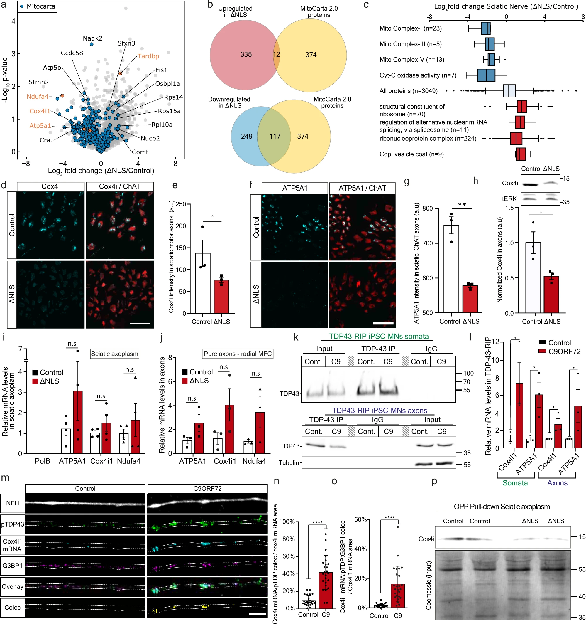
Yet another in-vitro and mice study was recently published. So even if there is eventually a positive outcome, it is many years in the future. Basically it tells about experimentation done on the axon of in-vitro and in a mice model. The conclusion is that TDP-43 (which is found in misfolded aggregates in >95% of ALS patients) impairs local mitochondria in neuromotor junctions (NMJ). enter image description here

However some questions could be asked: * The authors say TDP-43 in ALS plays an important role at NMJ. This is certainly NOT a mainstream statement: For most (but not all) scientists, ALS starts in the brain (upper motor neurons), not in the NMJs (lower motor neurons).

  • They use transgenic mice expressing the human TDP-43 lacking the nuclear-localization-signal (∆NLS). No wonder TDP-43 goes in weird places into the cell. To remind, once a protein is produced by the ribosomes and folded by the endoplasmic reticulum (ER), it is packaged and sent to its final destination (the nucleus for TDP-43) by the Golgi apparatus. If there is no NLS, then the newly protein is not sent somewhere, it just accumulates and moves at random pushed by the Brownian movement, but it is correctly folded because it went through the ER. This is not what scientists tell, for them in ALS the TDP-43 proteins do not enter in the ER, or the ER is dysfunctional, so they stay misfolded.

  • They confirm that misfolded TDP-43 is found in mitochondria, but this was first found by Gao and al at Case Western Uni in 2016.

  • When they reintroduce doxycycline in mice diet (a classic trick to switch ON/OFF a gene in a genetically modified mice), the TDP-43 got again its NLS and the mice health improved. Again this was shown in the past, specially by Gao and al in 2016 who designed a credible TDP-43 genetic therapy by adding a NLS signal to neuron cells. It remain to see in real life in humans if this would improve their health. After all "normal" TDP-43 got its NLS signal in the Golgi apparatus, so why this is not the case in ALS patients?

  • Something really interesting is that when they used TAT-fused peptide corresponding to residues 190-208 of G3BP1, a decrease in translation was reversed by axonal-exclusive application of (G3BP1 peptide) and mice health improved. This result mirrors other several experiments, yet this is done with peptides, not with costly genetic therapeis. This is something very low cost that could be easily tried in other labs in pre-clinical studies and may lead to a future therapy.

Contact the author

Advertisement


This book retraces the main achievements of ALS research over the last 30 years, presents the drugs under clinical trial, as well as ongoing research on future treatments likely to be able stop the disease in a few years and to provide a complete cure in a decade or two.
SGLT2 inhibitors, also called gliflozins, are a class of medications that alter essential physiology of the nephron. It inhibits reabsorption of glucose in the kidney and therefore lower blood sugar.

DPP-4 inhibitors increase incretin levels (GLP-1 and GIP), which inhibit glucagon release, which in turn increases insulin secretion, decreases gastric emptying, and decreases blood glucose levels.

The effects of sodium-glucose cotransporter 2 inhibitors (SGLT2I) and dipeptidyl peptidase-4 inhibitors (DPP4I) on new-onset cognitive dysfunction in type 2 diabetes mellitus remain unknown.

This study aimed to evaluate the effects of the two novel antidiabetic agents on cognitive dysfunction by comparing the rates of dementia between SGLT2I and DPP4I users.

This was a population-based cohort study of type 2 diabetes mellitus patients treated with SGLT2I and DPP4I between January 1, 2015 and December 31, 2019 in Hong Kong. Exclusion criteria were <1-month exposure or exposure to both medication classes, or prior diagnosis of dementia or major neurological/psychiatric diseases. * Primary outcomes were new-onset dementia, Alzheimer's, and Parkinson's. * Secondary outcomes were all-cause, cardiovascular, and cerebrovascular mortality.

Results: A total of 13,276 SGLT2I and 36,544 DPP4I users were studied, SGLT2I users had lower incidences of dementia , Alzheimer's, Parkinson's disease, all-cause, cerebrovascular, and cardiovascular mortality.

In conclusions the use of SGLT2I is associated with lower risks of dementia, Parkinson's disease, and cerebrovascular mortality compared with DPP4I use after 1:2 ratio propensity score matching.

Read the original article on Pubmed

Ropinirole extends survival by 28 weeks in an ALS clinical trial.

- Posted by admin in English

Some years ago there were great hopes in Pramipexole hydrochloride, a dopamine agonist. A phase III, multicenter, randomized, double-blind, placebo-controlled study of RPPX (EMPOWER) was conducted in ALS patients in the US, Canada, Australia, and Europe; however, regrettably, the results were clinically insignificant.

In 2018 Japanese scientists used induced pluripotent stem cell (iPSC) technology to generate stem and differentiated cells retaining the patients' full genetic information. They thus established a large number of in vitro cellular models of SALS. These models showed phenotypic differences in their pattern of neuronal degeneration, types of abnormal protein aggregates, cell death mechanisms, and onset and progression of these phenotypes in vitro among cases.

The researchers therefore developed a system for case clustering capable of subdividing these heterogeneous SALS models by their in vitro characteristics. They further evaluated multiple-phenotype rescue of these subclassified SALS models using agents selected from non-SOD1 FALS models, and identified ropinirole, a drug similar to Pramipexole, as a potential therapeutic candidate.

As a result, ropinirole hydrochloride was eventually selected. Therefore, the scientists wanted to explore the safety, tolerability and efficacy of ropinirole hydrochloride as an ALS treatment in this clinical trial.

Patient recruitment began in December 2018 and the scientists published their results on MedArXiv. Twenty one participants with Amyotrophic Lateral Sclerosis FRS-R scores greater than 2 points were randomly assigned using dynamic allocation to receive ropinirole or placebo for 24 weeks in the double-blind period.

enter image description here

Upon completion, participants could choose to participate in the following 24-week open-label active extension period. The primary outcomes were safety and tolerability. The secondary outcomes for the feasibility trial objective were the change in the ALS FRS-R) score, composite functional endpoint, combined assessment of function and survival, event-free survival, and time to [≤]50% forced vital capacity (blinded outcome assessment).

The participants were randomized into two groups (ropinirole group; n=14) and received ropinirole (n=13) or placebo (n=7) and the data of all participants were analysed using mixed-effects models for repeated measures together.

The incidence of gastrointestinal disorders (mainly, temporary mild nausea and diarrhoea) was high at 77% in the ropinirole group versus 14% in the placebo group). This is common in this type of clinical trial yet it is a major problem to keep long term adherence to the treatment.

Regarding the feasibility of verifying efficacy, there were no significant differences in the ALS FRS-R score and combined assessment of function and survival scores during the double-blind period for 6 months, while the participants in the ropinirole group had lived an additional 28 weeks without disease progression events compared with the placebo group at 12 months. It seems the effect of ropinorole became obvious only after 24 weeks. enter image description here

Ropinirole is thus found (by the authors) safe and tolerable for patients with ALS and this trial indicates feasibility for a subsequent large-scale trial.

Read the original article on medRxiv

Contact the author

Advertisement


This book retraces the main achievements of ALS research over the last 30 years, presents the drugs under clinical trial, as well as ongoing research on future treatments likely to be able stop the disease in a few years and to provide a complete cure in a decade or two.

This study investigated the criterion and construct validity of a novel, acoustic-based framework composed of five key components of motor control: Coordination, Consistency, Speed, Precision, and Rate.

Acoustic and kinematic analyses were performed on audio recordings from 22 subjects with amyotrophic lateral sclerosis during a sequential motion rate task. Perceptual analyses were completed by two licensed speech-language pathologists, who rated each subject's speech on the five framework components and their overall severity. Analytical and clinical validity were assessed by comparing performance on the acoustic features to their kinematic correlates and to clinician ratings of the five components, respectively.

Divergent validity of the acoustic-based framework was then assessed by comparing performance on each pair of acoustic features to determine whether the features represent distinct articulatory constructs. Bivariate correlations and partial correlations with severity as a covariate were conducted for each comparison.

Results revealed moderate-to-strong analytical validity for every acoustic feature, both with and without controlling for severity, and moderate-to-strong clinical validity for all acoustic features except Coordination, without controlling for severity. When severity was included as a covariate, the strong associations for Speed and Precision became weak.

Divergent validity was supported by weak-to-moderate pairwise associations between all acoustic features except Speed (second-formant [F2] slope of consonant transition) and Precision (between-consonant variability in F2 slope).

This study demonstrated that the acoustic-based framework has potential as an objective, valid, and clinically useful tool for profiling articulatory deficits in individuals with speech motor disorders. The findings also suggest that compared to clinician ratings, instrumental measures are more sensitive to subtle differences in articulatory function. With further research, this framework could provide more accurate and reliable characterizations of articulatory impairment, which may eventually increase clinical confidence in the diagnosis and treatment of patients with different articulatory phenotypes.

Read the original article on Pubmed

Contact the author

Advertisement


This book retraces the main achievements of ALS research over the last 30 years, presents the drugs under clinical trial, as well as ongoing research on future treatments likely to be able stop the disease in a few years and to provide a complete cure in a decade or two.

Mild Cognitive Impairment (MCI) is fraught with high false positive diagnostic errors. The high rate of false positive diagnosis hampers attempts to identify reliable and valid biomarkers for MCI.

Recent research suggests that aberrant functional neurocircuitries emerge prior to significant cognitive deficits. The aim of the present study was to examine this in clinically confirmed multi-domain amnestic-MCI (mdaMCI) using an established, multi-time point, methodology for minimizing false positive diagnosis.

Structural and resting-state functional MRI data were acquired in healthy controls (HC, n=24), clinically-confirmed multi-domain amnestic-MCI (mdaMCI, n=14) and mild Alzheimer's Dementia (mAD, n=6).

Group differences in cortical thickness, hippocampal volume and functional connectivity were investigated. Hippocampal subvolumes differentiated mAD from HC and mdaMCI.

Functional decoupling of fronto-temporal networks implicated in memory and executive function differentiated HC and mdaMCI.

Decreased functional connectivity in these networks was associated with poorer cognitive performance scores.

Preliminary findings suggest the large-scale decoupling of fronto-temporal networks associated with cognitive decline precedes measurable structural neurodegeneration in clinically confirmed MCI and may represent a potential biomarker for disease progression.

Read the original article on Pubmed


Please, help us continue to provide valuable information: