Parkinson's disease is a progressive neurodegenerative disease with no cure and few treatment options.

Photobiomodulation has been used successfully in animal models. enter image description here A new medRxiv preprint describes a proof-of-concept clinical study to assess the efficacy of photobiomodulation in Parkinson's disease, in order to inform on treatment regimens and outcome measures for a future randomized, placebo-controlled study.

Photobiomodulation therapy is the use of narrow wavelength bands of non-thermal light to modulate cellular responses. The main target of photobiomodulation is probably the cytochrome-C-oxidase, which absorbs red and near infrared light. Photobiomodulation therapy has a good safety profile.

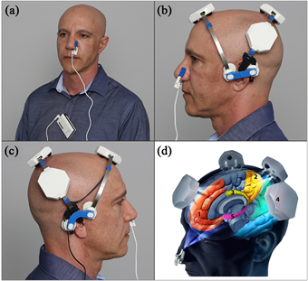
The photobiomodulation was administered transcranially with a VieLight Gamma device (4 LEDs, 240 joules), intranasally with a VieLight Gamma nasal device (1 LED, 15 joules), transdermally to the C1/C2 region of the neck and to the abdomen with an Irradia MID 2.5 laser device (4 laser diodes, 39.6 joules) or a MIDCARE laser device (2 diodes 39.6 joules).

All participants received the same dose of total energy from the photobiomodulation treatment throughout the study. The treatment protocol made it possible to not to use safety glasses. Participants' adherence to the treatment protocol was monitored by caregivers and reported during the final assessment.

The primary outcome measure was improvement timed up and go (TUG) as a measure of mobility. Secondary outcome measures were mobility, cognition, fine motor skill, micrographia and static balance. Quality of life outcome measures and patient-reported symptomatic changes, including depression, are reported separately.

Twelve participants with idiopathic Parkinson's disease were recruited. Six were randomly selected to start 12 weeks of transcranial, intranasal, cervical and abdominal photobiomodulation. The other 6 were put on a waiting list for 14 weeks before starting treatment. After the 12 week treatment period, all participants received photobiomodulation devices to continue treatment at home.

Participants were assessed for mobility, fine motor skills, balance, and cognition before the start of treatment, after 4 weeks of treatment, after 12 weeks of treatment and the end of the home treatment period.

Measures of mobility, cognition, dynamic balance and fine motor skills were significantly improved (p <0.05) with photobiomodulation treatment for 12 weeks and up to one year. Individual improvements varied, but many continued for up to a year with sustained home treatment. Improvements were maintained as long as treatment continued, up to a year in neurodegenerative disease where a decline is generally expected. No side effects of the treatment were observed.

The current study did not have a placebo arm to quantify the placebo effect, but the related Hawthorne effect was assessed. The Hawthorne effect occurs in response to participation in research. It happens when a patient is observed during a study and has been recognized as a confounding factor in the results of clinical trials in Parkinson's disease, for example depending on whether an evaluation is done openly or secretly.

In the present study, participants on the waiting list (group B) showed an improvement in outcome measures before the start of treatment, with some of these improvements being sufficient to qualify as IMID, thus demonstrating a measurable Hawthorne effect. The other possibility is that the participants improved due to a practice effect with repeated assessments.

In conclusion, photobiomodulation has been shown to be a safe and potentially effective treatment for a range of clinical signs and symptoms of Parkinson's disease. Home treatment for Parkinson's disease on its own or with the help of a caregiver can be an effective treatment option. The results of this study indicate that a large double-blind clinical trial of the application of this technology to Parkinson's disease is warranted.

TRIAL REGISTRATION: Australian New Zealand Clinical Trials Registry, registration. number: ACTRN12618000038291p, registered on 12/01/2018

Advertisement


This book retraces the main achievements of ALS research over the last 30 years, presents the drugs under clinical trial, as well as ongoing research on future treatments likely to be able stop the disease in a few years and to provide a complete cure in a decade or two.

In a new publication Gadd et al. apply blood-derived epigenetic signatures of lifestyle traits to matched blood and brain samples, uncovering variability in how well blood translates across brain regions and a relationship between smoking and the prefrontal cortex. These preliminary results contribute to understanding how lifestyle-related DNA methylation affects the brain. enter image description here These factors have been extensively linked with blood-based genome-wide DNA methylation, but it is unclear if the signatures from blood translate to the target tissue of interest-the brain. To investigate this, the authors apply blood-derived epigenetic predictors of four lifestyle traits to genome-wide DNA methylation from five post-mortem brain regions and the last blood sample prior to death in 14 individuals in the Lothian Birth Cohort 1936 (LBC1936).

This cohorte is a longitudinal study of healthy ageing in individuals who reside in Scotland. Participants completed a childhood intelligence test at age 11 years in 1947 and were then recruited for this cohort at the mean age of 70 years. They have been followed up approximately every 3–4 years (currently at the sixth wave), collecting a series of cognitive, physical, clinical and social data, along with blood donations that have been used for genetic, epigenetic, and proteomic measurement. Approximately 15% of individuals in the LBC1936 have consented to post-mortem tissue collection.

The LBC1936 group were born in 1936 and there are socioeconomic and cultural trends in their lifestyles that must be taken into consideration; many may have worked in factories or in shipbuilding yards, exposing them to high levels of respiratory pollutants and poorer socioeconomic status in this era was related to behaviours such as smoking. Though Scotland has a historic and sustained high prevalence of unhealthy lifestyle behaviours such as smoking and alcohol consumption, with a large proportion of the current population either overweight or obese, cohort profiling has found both the Generation Scotland and Lothian Birth Cohorts to be healthier and of higher socioeconomic status than the general population.

The majority of the 14 individuals within the brain bank subset either had been or were still smokers (86%) at the time of death, which was a higher proportion than in the reference group (46%). Most of the 14 individuals (86%) had high HDL cholesterol (> 1 mmol/l), drank alcohol weekly (92%) and had mean BMI of 25.5 kg/m2. Five of the individuals did not smoke.

Using these matched samples, the authors found that correlations between blood and brain DNA methylation scores for smoking, high-density lipoprotein cholesterol, alcohol and body mass index were highly variable across brain regions.

Smoking scores in the dorsolateral prefrontal cortex had the strongest correlations with smoking scores in blood and smoking behaviour. This was also the brain region which exhibited the largest correlations for DNA methylation at AhRR/site cg05575921 - which is the single strongest correlate of smoking in blood-in relation to blood and smoking behaviour. The aryl hydrocarbon receptor (AhRR) gene may act as a tumor suppressor but it is known that Dioxins and dioxin-like compounds are teratogens that exert their effects through the AhRR. This suggested a particular vulnerability to smoking-related differential methylation in this region.

The authors' work contributes to understanding how lifestyle factors affect the brain and suggest that lifestyle-related DNA methylation is likely to be both brain region dependent and in many cases poorly proxied for by blood. Though these pilot data provide a rarely-available opportunity for the comparison of methylation patterns across multiple brain regions and the blood, due to the limited sample size available authors' results must be considered as preliminary and should therefore be used as a basis for further investigation.

All R code used in this study is available with open access at the following Gitlab repository: https://gitlab.com/dannigadd/blood-brain-lifestyle-traits.

Advertisement


This book retraces the main achievements of ALS research over the last 30 years, presents the drugs under clinical trial, as well as ongoing research on future treatments likely to be able stop the disease in a few years and to provide a complete cure in a decade or two.

Un accident vasculaire cérébral (AVC) peut résulter en une incapacité modérée à sévère. La faiblesse musculaire après un AVC résulte de dommages aux fibres corticospinales qui relient le cortex moteur cérébral à la moelle épinière puis aux muscles. enter image description here

Les patients victimes d'un AVC souffrent souvent de troubles fonctionnels et ont besoin d'une rééducation. Une hémiparésie consiste en la paralysie partielle d’un des deux côtés (gauche ou droit) du corps.

Les scientifiques ne comprennent encore pas bien comment le corps se réorganise pour faire prendre en charge une partie des muscles par les nerfs qui n’ont pas été impactés par l’AVC.

Dans ce nouvel article, Urbin, Collinger et Wittenberg de l’université de Pittsburgh ont voulu quantifier le recrutement corticospinal des motoneurones spinaux servant les muscles intrinsèques de chaque main de treize patients atteints de déficience motrice de longue date secondaire à un accident vasculaire cérébral.

Pour cela ils ont effectué des expériences en utilisant la technique de triple stimulation. Les systèmes de stimulation du système nerveux par impulsions coordonnées sur la même fibre nerveuse permettent d'étudier la physiologie corticale du système moteur. La technique de triple stimulation délivre trois stimuli successifs: un premier dans le cortex moteur, un second sur le poignet, et un troisième sur le thorax (point d’Erb).

Lorsque dans les voies corticospinales intactes tous les neurones moteurs spinaux sont activés, les potentiels descendants entrent en collision avec les potentiels d'action ascendants résultant de la stimulation au niveau du poignet, et ils s'annulent mutuellement. Il en va différemment dans le cas d'un AVC. Dans ce cas, tous les potentiels d’action nerveux ascendants ne sont pas annulés et peuvent ainsi être détectés au niveau du thorax.

Une unité motrice est composée d'un neurone moteur et des fibres musculaires qu'il innerve. Des groupes d'unités motrices travaillent souvent ensemble pour coordonner les contractions d'un seul muscle. Quand une unité motrice devient défaillante (ici par ce qu’un neurone moteur est mort dans le cortex moteur à la suite d’un déficit en oxygène suite à l’AVC), les unités motrices voisinent peuvent prendre en charge une partie des connexions assurées par l’unité motrice défaillante. On appelle cela de la plasticité. Malgré tout cela se traduira par une maladresse et un manque de force.

La technique de triple stimulation permet de mieux quantifier la proportion d'unités motrices activées par stimulation magnétique transcrânienne.

Michael Urbin et ses collègues ont également examiné si les échecs d’activation musculaire pouvaient être inversés de manière transitoire en renforçant la connectivité synaptique via une stimulation non invasive.

Cette stimulation non invasive a entraîné de la plasticité, comme une augmentation du recrutement des motoneurones spinaux.

Les scientifiques ont aussi effectué des expériences de contrôle pour isoler le locus de plasticité. Ces expériences ont démontré une modulation chez les potentiels évoqués moteurs provoquée par la stimulation électrique du cortex moteur primaire mais pas par la taille ou la persistance de l'onde F. Une onde F est l'une des nombreuses réponses motrices qui peuvent suivre la réponse motrice directe induite par la stimulation électrique des nerfs moteurs périphériques ou mixtes.

Les ondes F sont évoquées quand des stimuli électriques sont appliqués à la surface de la peau au-dessus de la partie distale d'un nerf. Cette impulsion se déplace à la fois vers les fibres musculaires et vers le corps du neurone moteur dans la moelle épinière.

Ceci suggère que la plasticité motrice observée à la suite d’un AVC est médiée par une efficacité accrue des synapses neuronales cortico-motrices encore fonctionnelles. La modulation du recrutement est fortement associée à l'asymétrie initiale du recrutement et à la sévérité de la déficience.

Les résultats des scientifiques de l’université de Pittsburgh démontrent que les déficits de recrutement peuvent être partiellement et transitoirement inversés par la plasticité des synapses, en aval des circuits nerveux endommagés par un accident vasculaire cérébral.

Ils montrent que la plasticité des synapses dépendant de la synchronisation des pics entre les motoneurones corticospinaux et spinaux a inversé de manière transitoire les échecs de recrutement chez certains survivants.

Alzheimer’s disease has become the third leading cause of death.

Studies have provided candidate mechanisms for the neurodegeneration associated with Alzheimer’s disease, such as neuroinflammation, insulin resistance, and reduction in trophic support.

One complicating feature is that their etiology remains controversial, with many competing theories, such as “type 3 diabetes”, viral infection, misfolded or prions proteins, none of which has led to effective treatment.

The approach utilized in a new innovative trial departs sharply from traditional treatment strategies for MCI and Alzheimer’s disease, which have largely been monotherapeutic, monophasic, non-personalized, and blind, i.e., cause-independent, thus not targeted to the underlying drivers of the disease in each person, but rather to common downstream consequences and/or secondary drivers, such as amyloidosis.

In the field of oncology, a personalized, precision medicine approach, in which the presumptive molecular drivers of the disease process are targeted therapeutically, has improved outcomes in at least some studies.

However, this strategy has not been applied successfully to neurodegenerative diseases.

A recent BioRxiv article presents a proof-of-concept trial of precision medicine approach to Alzheimer’s disease.

Twenty-five patients with Alzheimer’s disease or mild cognitive impairment, ages 50-76, were recruited to three clinical sites and were evaluated for markers of inflammation, chronic infection, dysbiosis, insulin resistance, protein glycation, vascular disease, nocturnal hypoxemia, hormone insufficiency or dysregulation, nutrient deficiency, toxin or toxicant exposure (metals, organic toxicants, and biotoxins), genetic predisposition to cognitive decline, and other biochemical parameters associated with cognitive decline. Brain magnetic resonance imaging with volumetrics was performed at baseline and study conclusion.

Patients were treated for nine months with a personalized, precision medicine protocol that addressed each patient’s identified potentially contributory factors.

Diet was a plant-rich, high-fiber, mildly ketogenic diet, high in leafy greens and other nonstarchy vegetables(raw and cooked), high in unsaturated fats, with a fasting period of 12- 16 hours each night.

Exercise, both aerobic and strength training, was encouraged for at least 45 minutes per day, at least six days per week, and facilitated by the personal trainers. High-intensity interval training (HIIT) was recommended a minimum of twice per week.

Sleep hygiene was supported to ensure 7-8 hours of quality sleep per night, and all patients without known sleep apnea were tested over several nights using home sleep study devices.

For those patients with suboptimal hormonal status, bioidentical hormone replacement and appropriate supplements were provided to optimize sex hormone levels, neurosteroids, and thyroid medications as indicated for sub-optimal thyroid function.

Infectious agents associated with cognitive decline or systemic inflammation were identified and treated.

All outcome measures revealed improvement: statistically highly significant improvement in MoCA scores, CNS Vital Signs Neurocognitive Index, and AQ-C were documented. No serious adverse events were recorded.

On overall results support the notion that a precision medicine approach to the cognitive decline of Alzheimer’s disease and mild cognitive impairment may be an effective strategy, especially with continued optimization over time.

La progression des traits liés au vieillissement varie considérablement selon les individus, influencée par leur environnement, leur mode de vie et leur génétique. La notion de vieillissement biologique plutôt que chronologique, affirme que le vieillissement varie pas seulement le passage du temps mais aussi avec celui de l'organisme. Dans cette nouvelle étude, Michael Petr, Rafael de Cabo et ses collègues ont effectué des tests physiologiques et fonctionnels tout au long de la durée de vie des souris mâles de type C57BL/6N. La souris C57BL/6, qui peut vivre jusqu'à 4 ans, présente de nombreuses caractéristiques inhabituelles pour une souris de laboratoire: elle est exceptionnellement sensible à la douleur et au froid, et les médicaments analgésiques y sont moins efficaces. Contrairement à la plupart des souches de souris, ce type de souris est plus sensible aux addictions, à l'athérosclérose et la perte auditive avec l'âge.

En parallèle des tests fonctionnels, les scientifiques ont réalisés des analyses métabolomiques du sérum, du cerveau, du foie, du cœur et des muscles squelettiques afin d'identifier les signatures associées à l'âge. enter image description here Les auteurs ont effectué une caractérisation approfondie de ces différences parmi trois groupes d'animaux: Jeunes, adultes et vieux.

Cette analyse a démontré que: (i) la diminution de la vitesse de marche est un biomarqueur fonctionnel majeur du vieillissement chez la souris; (ii) la détérioration de l'activité locomotrice est associée à une augmentation spectaculaire du coût énergétique de l'activité physique à partir de l'âge de 19 mois environ, et s'accompagne d'une diminution soutenue de la capacité de travail;

Pour explorer davantage les changements moléculaires qui peuvent contribuer ou résulter de ces modifications, les scientifiques ont effectué une analyse métabolomique dans un certain nombre de tissus métaboliques clés et ont montré que: (i) différents organes remodèlent leur métabolisme en réponse à des demandes fonctionnelles spécifiques, par exemple l'approvisionnement en énergie et la désintoxication. (ii) l'épuisement du glucose, du 3-HB et du glycerol dans le sérum des souris agées montre une contribution réduite du foie et du tissu adipeux, aux autres organes; (ii) le vieillissement chez la souris favorise la modulation à la hausse du métabolisme du glucose dans les muscles cardiaques et squelettiques ainsi que dans le foie, où la gluconéogenèse et le cycle de l'urée sont également améliorés. Il y a un schéma similaire mais moins prononcé dans le cerveau.

Les auteurs ont ensuite évalué les associations entre les métabolites et les paramètres phénotypiques en sélectionnant les 24 métabolites les plus représentatifs indépendamment de l'âge et de l'organe. Les principaux résultats ont montré que la variance du coût énergétique et du rapport d'échange respiratoire peut être expliquée par un modèle distinct de remodelage métabolique dans le foie (par exemple, métabolisme mixte du glucose, des lipides et des acides aminés), des muscles cardiaques et squelettiques (par exemple, glucose et lipides), et cerveau (mélange d'acides aminés en plus du glucose et du catabolisme corporel cétonique).

L'augmentation de la demande énergétique a suscité une utilisation de plus en plus mixte des substrats (glucose, lipides, acides aminés) conduisant à une augmentation du stress oxydatif dans les organes qui ont une capacité antioxydante plus faible par rapport au cœur et au foie.

L'accumulation de graisse viscérale est associée à la résistance à l'insuline, tandis que la graisse sous-cutanée joue un rôle dans la réduction des niveaux d'insuline et l'amélioration de la sensibilité à l'insuline. En effet, l'augmentation du pourcentage de graisse corporelle des souris jeunes aux souris adultes coïncidait avec un rapport graisse sous-cutanée / viscérale plus élevé, ce qui, à son tour, était associé à une baisse significative des taux d'insuline circulante et de la résistance à l'insuline.

Les auteurs supposent que les vieux animaux, peuvent moins compter sur l'oxydation des graisses (en utilisant les graisses stockées) et plus sur l'utilisation moins efficace des glucides pour les besoins énergétiques.

Cette utilisation de plus en plus mixte du glucose et des lipides a conduit à une tendance vers une plus grande abondance de méthionine sulfoxyde et une déplétion en glutathion en réponse à une augmentation du taux d'échange respiratoire dans le cœur et le foie, alors que le cerveau avait nettement moins de méthionine sulfoxyde, mais une plus grande accumulation de méthionine et de nicotinamide par rapport à glutathion. Ce schéma est cohérent avec un stress oxydatif systémique et une capacité antioxydante plus faible.

Ensemble, ces données suggèrent qu'à mesure que les souris vieillissent, des organes tels que le foie et le cœur, qui sont exposés à un stress oxydatif plus élevé en raison de leur fonction (détoxification du foie et génération d'énergie dans le cœur), remodèlent leur métabolisme vers une expression plus élevée / l'activité des voies métaboliques liées à l'oxydoréduction, par exemple, le pentose phosphate, la récupération NAD + et la transsulfuration. À l'inverse, le muscle squelettique et le cerveau ne semblent pas capables de remodeler ces voies, devenant ainsi plus vulnérables à un stress oxydatif accru avec le vieillissement. Ce modèle métabolique est en accord avec l'idée de différents taux de vieillissement parmi les organes.

The progression of aging-related traits varies considerably among individuals, influenced by their environment, lifestyle and genetics. The notion of biological rather than chronological aging states that aging varies not only with the passage of time but also with that of the organism. In this new study, Michael Petr, Rafael de Cabo and their colleagues performed physiological and functional tests throughout the lifespan of male C57BL/6N mice. The C57BL/6 mouse, which can live up to 4 years, exhibits many characteristics unusual for a laboratory mouse: it is exceptionally sensitive to pain and cold, and analgesic drugs are less effective in it. Unlike most strains of mice, this type of mouse is more susceptible to addiction, atherosclerosis and hearing loss with age.

In parallel with the functional tests, the scientists performed metabolomic analyzes of serum, brain, liver, heart and skeletal muscles in order to identify the signatures associated with age. enter image description here The authors performed an in-depth characterization of these differences among three groups of animals: Young, Adult and Old.

This analysis showed that: (i) decreased walking speed is a major functional biomarker of aging in mice; (ii) the deterioration of locomotor activity is associated with a dramatic increase in the energy cost of physical activity from the age of about 19 months, and is accompanied by a sustained decrease in working capacity;

To further explore the molecular changes that may contribute to or result from these changes, scientists performed metabolomic analysis in a number of key metabolic tissues and showed that: (i) different organs reshape their metabolism in response to specific functional demands, eg energy supply and detoxification. (ii) depletion of glucose, 3-HB and glycerol in the serum of elderly mice shows reduced contribution from liver and adipose tissue to other organs; (ii) aging in mice promotes upward modulation of glucose metabolism in cardiac and skeletal muscles as well as in the liver, where gluconeogenesis and urea cycling are also enhanced. There is a similar but less pronounced pattern in the brain.

The authors then assessed the associations between metabolites and phenotypic parameters by selecting the 24 most representative metabolites regardless of age and organ. The main results showed that the variance of the energy cost and the respiratory exchange ratio can be explained by a distinct model of metabolic remodeling in the liver (eg, mixed metabolism of glucose, lipids and amino acids), muscles cardiac and skeletal (eg, glucose and lipids), and brain (mixture of amino acids in addition to glucose and ketone body catabolism).

The increase in energy demand has prompted an increasingly mixed use of substrates (glucose, lipids, amino acids) leading to an increase in oxidative stress in organs which have a lower antioxidant capacity compared to the heart and the liver. .

The accumulation of visceral fat is associated with insulin resistance, while subcutaneous fat plays a role in lowering insulin levels and improving insulin sensitivity. Indeed, the increase in the percentage of body fat from young mice to adult mice coincided with a higher subcutaneous / visceral fat ratio, which, in turn, was associated with a significant decrease in circulating insulin levels and insulin resistance.

The authors assume that older animals can rely less on fat oxidation (using stored fat) and more on less efficient use of carbohydrates for energy needs.

This increasingly mixed use of glucose and lipids has led to a trend towards greater abundance of methionine sulfoxide and depletion of glutathione in response to an increased rate of respiratory exchange in the heart and liver, while the brain had significantly less methionine sulfoxide, but a greater accumulation of methionine and nicotinamide compared to glutathione. This pattern is consistent with systemic oxidative stress and lower antioxidant capacity.

Taken together, these data suggest that as mice age, organs such as the liver and heart, which are exposed to higher oxidative stress due to their function (detoxifying the liver and generating energy in the heart). ), remodel their metabolism towards higher expression / activity of redox-related metabolic pathways, eg, pentose phosphate, NAD + recovery and transsulfurization. Conversely, skeletal muscle and the brain do not appear to be able to reshape these pathways, thus becoming more vulnerable to increased oxidative stress with aging. This metabolic model is consistent with the idea of different rates of aging among organs.

Advertisement


This book retraces the main achievements of ALS research over the last 30 years, presents the drugs under clinical trial, as well as ongoing research on future treatments likely to be able stop the disease in a few years and to provide a complete cure in a decade or two.

Another attempt at finding an ALS pathogen is unsuccessful

- Posted by admin in English

As various pathogens have been reported in the blood, cerebrospinal fluid, and central nervous system of patients with ALS, a few scientific publications have suggested that infectious agents may play a role in neurodegenerative diseases, but these agents have never been identified.

In a new scientific publication, the authors report they have found an unidentified virus like signature in 120 whole blood RNA samples, in ALS patient as well in controls samples. However when they used other public databases they where unable to find this viral signature, so little have been learned.

The search for pathogens using sequencing data from blood samples in patients with ALS has already been done, but as sequencing techniques can only read tiny fragments of DNA at the same time (the "reads"), they must recourse to reference genomes to succeed in a reconstitution, and this reconstitution does not allow the discovery of genomes are not included in the reference bases.

To complicate matters further, the readings belong to the patient and not to an hypothetical microbe or virus.

So when Melnick, Prudencio and his colleagues at Boulder and Jacksonville set out to design a new sequencing pipeline they made sure it does not ignore "reads" that cannot be aligned with a known genome.

The authors developed a bioinformatics pipeline that identifies microbial sequences in mammalian RNA-seq data, including sequences without significant nucleotide similarity results in GenBank.

They opted for a de novo assembly of unmapped reads into contigs, followed by aligning unmapped reads to these contigs for quantification. The code used in this manuscript is available at https://github.com/Senorelegans/MysteryMiner

A total of 120 whole blood RNA samples were initially used. It included 30 healthy controls (from the general population who do not have blood relatives with ALS), 30 pre-symptomatic C9ORF72 mutant carriers, 30 symptomatic cases of C9ORF72 ALS, and 30 cases symptomatic C9ORF72 negative ALS.

The efficiency of this pipeline has been tested by the authors on public RNA-seq data. The scientists then applied this pipeline to a new RNA-seq dataset generated from a cohort of 120 samples from patients and controls with amyotrophic lateral sclerosis (ALS), and identified sequences corresponding to bacteria and known viruses, as well as new virus-like sequences.

The complete dataset contains 8.64 X 109,406 combined reads. About 2.7% (2.34 X 10 ^ 8) of the reads did not match the human genome. From these non-host reads, 2,976,988 contigs were assembled and 17,047 BLASTN (regular biome) contigs were identified. A total of 25,815 contigs did not match by BLASTN and after filtering they identified 2,980 dark biome contigs (identified by BLASTX) and 859 double dark biome contigs (no BLASTX or BLASTN hit).

In the dark biome contigs, Melnick and his colleagues noted many contigs with a region of protein sequence similar to the RNA-dependent RNA polymerase (RdRP) of several RNA viruses, showing the greatest similarity to the virus. velvet tobacco marbling. This was present in the control as well.

RdRP is an essential protein encoded in the genomes of all viruses containing RNA without a DNA stage, that is to say RNA viruses including SARS-CoV-2.

To validate that this virus-like sequence was not a contig assembly artifact or a contaminant introduced during library construction or sequencing, the authors used RT-PCR of the original patient samples to demonstrate that this sequence was present in positive samples identified by RNA-seq analysis and not detectable in negative samples.

The scientists then investigated whether similar results would be obtained from other ALS data sets. To this end, they examined five other publicly available ALS datasets.

However, they found no statistically significant difference between samples from patients with ALS and control samples for virus / bacteria genus / species in normal / dark biome for any of the remaining ALS datasets.


Please, help us continue to provide valuable information: