The insulin signaling pathway plays a crucial role in regulating the growth and metabolism of neurons. Deregulation of IGF-R signaling has been linked to a variety of neurodegenerative diseases such as Alzheimer's, Parkinson's and Huntington's disease.

However, the role of insulin signaling in C9orf72 ALS / FTD is not yet clear. A positive correlation of the incidence of ALS with early-onset type 1 diabetes has been reported, and a decrease in insulin and IGF-1 in the blood and cerebrospinal fluid of patients with ALS, although the relevance of these findings to disease progression is unclear.

Intrathecal administration of IGF-1 improved motor performance, delayed disease onset, and prolonged survival in the SOD1G93A mouse model of ALS. However, three clinical trials of IGF-1 administered subcutaneously in ALS have reported conflicting results. The contradictory result of these trials may be due to insufficient administration of drugs to the brain and spinal cord and the fact that ALS has heterogeneous genetic risk factors.

Atilano, Isaacs, Partridge, and colleagues have shown in a recent article that insulin/IGF signaling is reduced in C9orf72 fly models using adult brain RNA sequencing. They further demonstrated that activation of insulin/IGF signaling can attenuate several neurodegenerative phenotypes in flies expressing expanded G4C2 repeats or the toxic dipeptide repeat protein poly-GR.

Poly-GR levels are reduced when components of the insulin/IGF signaling pathway are genetically activated in diseased flies, suggesting a rescue mechanism. This effect on poly-GR levels was confirmed in a mammalian cell model. Modulation of insulin signaling in mammalian cells also lowers poly-GR levels. Remarkably, the systemic injection of insulin improves the survival of flies expressing G4C2 repeats. Their data suggest that modulation of insulin/IGF signaling may be an effective therapeutic approach against C9orf72 ALS/FTD.

PTEN acts as a tumor suppressor gene through the action of its protein phosphatase product. This phosphatase is involved in the regulation of the cell cycle, preventing cells from growing and dividing too quickly. It is a target of many anticancer drugs. Pten reduction has also been reported to reduce the toxicity of C9orf72 repeats expressed in a mammalian cell line, again in agreement with the results described by scientists and the potential therapeutic benefit of modulating this pathway. It is also interesting to note that the process of brain aging has been linked to decreased insulin signaling as well as impaired insulin binding, which may partly explain why aging is a problem. risk factor for the disease.

The authors found that intrathoracic insulin administration prolonged the survival of flies expressing G4C2 repeats. Although robust, the lengthening of the lifespan was relatively modest, which could be explained by the transient nature of the insulin treatment. Insulin and IGF-1 ligands have already been tested in neurodegenerative diseases.

Overall, the researchers' study suggests that modulation of the insulin / IGF signaling pathway could be an effective therapeutic intervention against hexanucleotide repeat extension associated with C9orf72 neurodegenerative diseases, with InR being a genetic modifier. It will be interesting in the future to study the need for downstream effectors of insulin signaling in the rescue of toxicity. It is important to note that in the Drosophila, there is a single insulin-like system that has the dual function of insulin / IGF signaling; thus, the mechanism of toxicity described in the researchers' model could also be linked to IGFs. Therefore, it will be important to test whether treatment with insulin or IGF can save the survival of other C9orf72 ALS / FTD vertebrate model organisms.

Advertisement


This book retraces the main achievements of ALS research over the last 30 years, presents the drugs under clinical trial, as well as ongoing research on future treatments likely to be able stop the disease in a few years and to provide a complete cure in a decade or two.

An ALS therapy poses a moral dilemma to Biogen

- Posted by admin in English

It was found in 2009 that toxicity of SOD1 is secondary to a gain of toxic function rather than a loss of enzymatic function of the SOD1 enzyme, thus reducing levels of the mutant protein was predicted to slow progression in SOD1-linked ALS. Tofersen (formerly known as BIIB067), is an antisense oligonucleotide designed for adults with a confirmed superoxide dismutase 1 (SOD1) genetic mutation, a subtype of familial ALS that makes up 2 percent of all ALS cases. 

As reducing levels of the mutant protein is predicted to slow progression in SOD1-linked ALS, this antisense oligonucleotide mediates the degradation of superoxide dismutase 1 (SOD1) messenger RNA. As there is less SOD1 mRNA, there is less production of the SOD1 protein. This is not without risks in itself, SOD1’s role is to protect against anti-oxidants which are the normal by-product of cellular respiration.

Tofersen was developed in a long time partnership between Biogen and Ionis Pharmaceuticals.  With the time doses went from 3mg up to 100mg. They conducted a phase 1-2 ascending-dose trial evaluating Tofersen in adults with ALS due to SOD1 mutations. In each dose cohort (20, 40, 60, or 100 mg), participants were randomly assigned in a 3:1 ratio to receive five doses of Tofersen or placebo, administered intrathecally for 12 weeks.  The results were presented in July 2020.

The results were encouraging, but as for any ALS trial they are anything but stellar. Lumbar puncture-related adverse events were observed in most participants. Among participants who received Tofersen, one died from pulmonary embolus on day 137, and one from respiratory failure on day 152; one participant in the placebo group died from respiratory failure on day 52.

48 participants received all five planned doses. The technical indicators where better for participants having the higher doses. Patients taking the highest dose of Tofersen saw their ALS Functional Rating Scale score decrease by an average of 1.2 points, while the placebo group saw their score decrease by an average of 5.63 points.

Toby Ferguson the principal investigator acknowledged: “What we saw is a decline in ALS function, breathing and strength that was much greater in the placebo groups than in the treated groups. That said, if I were critical, I could poke holes in any one piece of those data, but we also have the data that SOD1 is being reduced.”

ALS is a difficult field in which drugs that show early promise can turn out to offer no benefit, or pose “unjustified risks” to patients in later studies. Biogen knows this as well as anyone, having seen its last ALS effort, dexpramipexole, fail in phase 3. Since then, the company has switched its focus from drugs with specific mechanisms, like anti-inflammatories, to medicines that target genetic mutations. Biogen strategy is  to learn more about the biology of the disease from looking at genetically defined populations (SOD1, C9orf72, ATXN2, XPO1) and then apply those findings to the broader population.

Despite those mixed results and as for other ALS drugs (Nurown, Cu(II)ATSM, Masitinib, AMX0035), there is an online petition for a compassionate access. Nearly 70,000 people have signed this online petition asking Biogen to provide an experimental ALS drug to a patient requesting it under compassionate use, but Biogen disagrees.

At the heart of Biogen’s denial is the question of broader access. The fastest way to get Tofersen to as many patients as possible would be regulatory approval, wrote Biogen chief medical officer, Maha Radhakrishnan, M.D., in a March 17 letter to an ALS patient, Stockman Mauriello that she shared with WCNC. That, of course, requires the completion of clinical trials.

“Providing individual access to Tofersen at this time could jeopardize access to Tofersen for hundreds of SOD1-ALS patients by impeding our ability to complete the study that will determine whether Tofersen is efficacious and safe and to seek subsequent regulatory approvals as quickly as possible,” Radhakrishnan wrote.

It's a moral position, it would not be fair to study participants who were randomized to placebo if Biogen were to offer the drug to patients not participating in the study.

However Biogen will provide early access to Tofersen only after study participants are no longer taking placebo.

“We cannot overlook these patients when considering questions of broader access, and cannot keep them on placebo while at the same time offering Tofersen to those outside of our study,” Sandrock wrote. “Offering Tofersen outside of the study would risk failing to complete the study and risk failing to obtain access for all SOD1-ALS patients".

“These patients agreed to participate in our study acknowledging the risk that they may not receive Tofersen, and with hope that Tofersen could be shown to work and be approved for all patients,” Sandrock wrote.

It has been clear for a while that anti-amyloid antibodies can sweep plaque from the brain, but until now the question of whether this slows cognitive decline has remained hotly contended. Despite some positive signals from four such antibodies, the data have been messy and hard to interpret.

In a Phase 2 trial, donanemab completely cleared plaque in two-thirds of participants. At the 15th International Conference on Alzheimer’s and Parkinson’s Diseases, held virtually March 10–14, Mark Mintun of Eli Lilly & Company presented the Phase 2 trial of the company’s anti-amyloid antibody donanemab.

This is the first anti-amyloid AD drug to meet a clinical endpoint in a Phase 2 trial.  While the drug met its primary endpoint, participants did not get better. Even so, donanemab slowed their decline by an average of 32 percent on a combined cognitive and functional measure. The treatment appeared to nudge down tangles, and worked best at low tangle loads.

The donanemab Phase 2 study offers some of the most compelling evidence to date that amyloid reduction can slow clinical decline, albeit modestly, in early stages of AD. In addition to the encouraging results, there are two unique features of this trial that move the field forward.

First is the decision by design to reduce dose or switch to placebo once patients are in the amyloid-negative range on PET. The discontinued dosing once the participant’s amyloid level normalizes may suggest that, if replicated, there will be not need to treat patients continuously. Doctors may be able to lower their patients amyloid levels, monitor them and, if the levels rise, re-dose. 

The second decision is the selection of patients with intermediate tau aggregates on PET. It is interesting that the overall clinical benefit was driven by the patients in the lowest tau PET tertile. This suggests that the primary role of amyloid-lowering therapies may be in patients in whom tau is not yet widespread, most of whom will be in the preclinical or very earliest clinical stage.

Advertisement


This book retraces the main achievements of ALS research over the last 30 years, presents the drugs under clinical trial, as well as ongoing research on future treatments likely to be able stop the disease in a few years and to provide a complete cure in a decade or two.

Parkinson's disease is a common neurodegenerative disease. Although the exact etiology and natural course of this disease have yet to be fully clarified, numerous system-level processes and dysfunctions, have been implicated in the pathogenesis of Parkinson's disease.

Lipid droplets are highly dynamic organelles that emerge from the endoplasmic reticulum (ER) membrane. lipid droplets is involved in fatty acid storage and participate in many diseases. For example, myeloid cells, including macrophages, leukocytes, and eosinophils, form lipid droplets in response to inflammation and stress. enter image description here

Normally, intracellular lipid droplets are degraded in lysosomes and deliver fatty acids to mitochondria for their consumption as an alternative energy source during periods of nutrient depletion. However, neurons have a low capacity for mitochondrial fatty acid consumption for energy production. This characteristic makes neurons particularly sensitive to lipid droplet accumulation and peroxidation.

Furthermore, the accumulation of lipid droplets enhances the fatty acid oxidation rate and imposes persistent pressure on the mitochondrial electron transport chain. Lipid overload also leads to ROS production by oxidative enzymes. Emerging evidence indicates that unexpected lipid droplet deposition and peroxidation can accelerate organelle stress and plays a crucial role in the pathogenesis of neurodegenerative diseases.

In a previous study, Xiaojuan Han and colleagues found that kaempferol, a natural flavonoid small molecule, exhibited neuroprotective effects on mice with drug-induced Parkinson's disease.

In the current study, the authors showed in-vitro that kaempferol protected against tyrosine hydroxylase neuronal loss and behavioral deficits in methyl-4-phenyl-1,2,3,6-tetrahydropyridine (MPTP) induced Parkinson disease mice, accompanied by reduced lipid oxidative stress in the substantia nigra pars compacta.

Kaempferol-rich food has been linked to a decrease in the risk of developing some types of cancers and cardiovascular disease. Quercetin and kaempferol are among the most ubiquitous polyphenols in fruit and vegetables.

Kaempferol is a chemical produced by Kaempferia galanga, one of four plants called galangal. The extract of Kaempferia galanga causes central nervous system depression, a decrease in motor activity, and a decrease in respiratory rate.

The mice were treated with kaempferol. The rotarod performance time was markedly reduced in MPTP-treated mice, and this effect was prevented by kaempferol. kaempferol also restored the behavioral deficits induced by MPTP, as indicated by the reductions in the turning time and total time in the pole test, without affecting the body weights of the mice.

MPP+, the active metabolite of MPTP, inhibits mitochondrial complex enzymes and causes the cell death that is directly associated with PD. In cultured neuronal cells, kaempferol exhibited a relatively safe concentration range and significantly suppressed lipid droplet accumulation and cellular apoptosis induced by MPP.

Further studies indicated that the protective effect of kaempferol was dependent on autophagy, specifically lipophagy. Critically, kaempferol promoted autophagy to mediate lipid droplet degradation in lysosomes, which then alleviated lipid deposition and peroxidation and the resulting mitochondrial damage, consequently reducing neuronal death.

Furthermore a genetic knockdown abolished the neuroprotective effects of kaempferol against lipid oxidation in Parkinson disease mice.

This work demonstrates that kaempferol prevents dopaminergic neuronal degeneration in Parkinson disease via the inhibition of lipid peroxidation-mediated mitochondrial damage by promoting lipophagy and provides a potential novel therapeutic strategy for Parkinson disease and related NDDs.

A new article by Qaisar, Qayum and Muhammad made me think about links between severe forms of asthma and some ALS forms. Indeed this has already been questioned by scientists. There are only a few studies addressing skeletal muscle function in patients with moderate to severe asthma. enter image description here

Asthma is the result of chronic inflammation of the conducting zone of the airways (most especially the bronchi and bronchioles), which subsequently results in increased contractability of the surrounding smooth muscles. This among other factors leads to bouts of narrowing of the airway and the classic symptoms of wheezing. The narrowing is typically reversible with or without treatment. Occasionally the airways themselves change.

Patients with asthma have diminished activity patterns, leading to deleterious physiologic alterations and ultimately impaired functional capacity. Moreover, they may use corticosteroids during periods of disease exacerbation.

Yet patients with asthma who use systemic corticosteroids present a decrease in respiratory muscle strength. The use of high doses of glucocorticoids causes atrophy and loss of muscle strength, a condition known as steroid myopathy.

An effective pharmacological intervention in asthma remains elusive, partly because molecular mechanisms dictating muscle decline in asthma are not known.

The authors of this new article about the relation between asthma and muscle wasting, investigated the potential contribution of skeletal muscle sarcoplasmic reticulum Ca2+ ATPase (SERCA) to muscle atrophy and weakness in asthmatic patients.

SERCA resides in the sarcoplasmic reticulum within myocytes. The sarcoplasmic reticulum is a membrane-bound structure found within muscle cells that is similar to the endoplasmic reticulum in other cells. The main function of the sarcoplasmic reticulum is to store calcium ions (Ca2+). Calcium ion levels are kept relatively constant, with the concentration of calcium ions within a cell being 10,000 times smaller than the concentration of calcium ions outside the cell.[

SERCA transfers Ca2+ from the cytosol of the cell to the lumen of the sarcoplasmic reticulum. This uses energy from ATP hydrolysis during muscle relaxation.In addition to its calcium-transporting functions, SERCA also generates heat in brown adipose tissue and in skeletal muscles

Quadriceps muscle biopsies were taken from 58 to 72 years old male patients with mild and advanced asthma and the SERCA activity was analyzed in association with cellular redox environment and myonuclear domain size.

Maximal SERCA activity was reduced in skeletal muscles of mild and advanced asthmatics and was associated with reduced expression of SERCA protein and upregulation of sarcolipin, a SERCA inhibitory lipoprotein.

Sarcolipin is an important mediator of muscle based non shivering thermogenesis. It causes the sarcoplasmic reticulum Ca2+-ATPases to stop pumping Ca2+ ions but continue futilely hydrolysing ATP, thus releasing the energy as heat. Sarcolipin mediated heat production is very important for many organisms to maintain a warm body. In mammals thermogenesis by skeletal muscles is complemented by thermogenesis in the brown adipose tissue.

The authors also found downregulation of Ca2+ release protein calstabin and upregulation of Ca2+ buffer, calsequestrin in skeletal muscles of asthmatic patients. The atrophic single muscle fibers had smaller cytoplasmic domains per myonucleus possibly indicating the reduced transcriptional reserves of individual myonuclei. Plasma periostin and C-terminal Agrin Fragment 22 (CAF22) levels were significantly elevated in asthmatic patients and showed a strong correlation with hand-grip strength. These changes were accompanied by substantially elevated markers of global oxidative stress including lipid peroxidation and mitochondrial ROS production.

Agrin, a synaptically located protein, is a key player during initial formation and maintenance of neuromuscularjunctions (NMJ). During development, nerve cells approach muscle fibers and establish synaptic contacts defined as NMJ. Initially, the NMJ is small and weak, but if the contact is successful the connection is maintained and reinforced. Agrin induces and stabilizes clusters of AChRs at the NMJ, promotes synaptic maturation, and maintains the mature state of the NMJ.

C-terminal Agrin Fragment has been proposed as a novel biomarker for sarcopenia originating from the degeneration of the neuromuscular junctions. Manipulation of the agrin signaling path may therefore be a promising way to correct neuromuscular defects.

During development in humans, the growing end of motor neuron axons secrete agrin. When secreted, agrin binds to several receptors on the surface of skeletal muscle. The receptor which appears to be required for the formation of the neuromuscular junction (NMJ) is called the MuSK receptor (Muscle specific kinase). Elevation of MuSK signaling has improved motor performance and delayed denervation in ALS mice (Perez-Garcia and Burden, 2012).

Taken together, the scientists' data suggest that muscle weakness and atrophy in asthma is in part driven by SERCA dysfunction and oxidative stress. The data propose SERCA dysfunction as a therapeutic intervention to address muscle decline in asthma.

You can write me at contact at padiracinnovation.org

Advertisement


This book retraces the main achievements of ALS research over the last 30 years, presents the drugs under clinical trial, as well as ongoing research on future treatments likely to be able stop the disease in a few years and to provide a complete cure in a decade or two.

Nearly 3 out of 4 cancer patients undergoing chemotherapy suffer from loss of memory, attention or concentration. This range of cognitive deficiencies referred to as chemotherapy-induced cognitive impairment, chemobrain or chemofog severely hampers quality of life of patients undergoing treatment. Advanced neuroimaging analyses have identified structural white and gray matter damage following chemotherapy in patients treated for various types of cancer. Cognitive deficits associated with platinum-based therapeutics such as cisplatin have been observed for 5-10 years post-diagnosis. However, no United States Food and Drug Administration-approved therapeutic interventions are available to date.

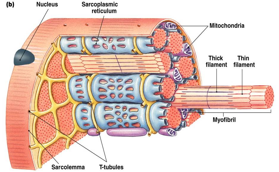
Mitochondrial dysfunction, characterized by abnormal morphology, impaired bioenergetics, altered mitochondrial dynamics and mitochondrial DNA mutations, has emerged as an underlying mechanism of several pathologies, including neurodegenerative diseases, cerebral and cardiac ischemia , traumatic brain injury, spinal cord injury, cancer, and chemotherapy-induced cognitive impairment and peripheral neuropathy. Following cerebral ischemia, astrocytes donate their healthy mitochondria to ischemic neurons with dysfunctional mitochondria to maintain adequate mitochondrial function and survival.

Studies in the 1930s already showed that nasally administered bacteria can cross the nasal epithelium within minutes, indicating a paracellular route of entry.

In a recent study, Jenolyn F. Alexander, Cobi J. Heijnen and colleagues demonstrated that nasal administration of mesenchymal stem cells restores cisplatin-induced cognitive impairment in mice and their data indicated that these mesenchymal stem cells act by repairing neuronal mitochondrial damage. enter image description here

It appeared that autologous mesenchymal stem cells work as well as human mesenchymal stem cells in their mouse model. The concept of mitochondrial administration is also being considered the treatment of Parkinson's disease, cerebral and cardiac ischemia, cancer, diabetic nephropathy and spinal cord injuries and some clinical trials for evaluating the safety and efficacy of isolated mitochondria based therapies have also recently commenced.

These findings indicate that cisplatin-induced accumulation of mitochondrial p53 is the cause of the damage to brain mitochondria that leads to cognitive deficits in response to treatment with this chemotherapeutic. The authors recently showed that astrocytes donate their healthy mitochondria and rescue primary cortical neurons damaged by cisplatin in vitro.

Based on their above-mentioned findings that mitochondrial deficits underlie cisplatin-induced neuronal damage and cognitive deficits, and that healthy mitochondria can be taken up by damaged neurons, the scientists hypothesized that isolated mitochondria from healthy mesenchymal stem cells can be used to resolve cisplatin-induced cognitive deficits and the associated structural damage. This would provide an advantage from the perspective of safety because the scientists do not need donation of allogeneic intact cells.

Mice were intraperitoneally injected with cisplatin at 2.3 mg/kg for 5 consecutive days, followed by 5 days of rest and another 5 days of cisplatin injection. The two administrations of mitochondria on mice delivered nasally, completely restored within two weeks mice's executive functioning, spatial recognition and working memory previously impaired by cisplatin treatment. The entry of mesenchymal stem cells into the brain is facilitated by pretreatment of the nasal cavity with hyaluronidase. The scientists maintained the mitochondria in calcium-free mitochondrial respiration buffer until ready for nasal delivery when they were transferred to calcium-free PBS.

Within 30 min of nasal administration, the mitochondria were detectable in the meninges where they were predominantly internalized by macrophages. Within this 30 min, the mitochondria also arrived at the ventricles and choroid plexus, gaining access to the brain.

3 h after delivery, mitochondria were found distributed along the rostral migratory stream where they were internalized by GFAP+ cells. By this time the nasally administered mitochondria also reached the hippocampus. Evidently, these mitochondria that had entered the meninges and brain completely restored the cisplatin-induced white matter damage in the cingulate cortex, synaptic loss in the hippocampus, and the compromised synaptosome membrane integrity and structural mitochondrial defects in synaptosomes as observed 35 days after the second mitochondrial donation.

The scientists used mitochondria isolated from human mesenchymal stem cells in their mouse model for the primary purpose of being able to trace the administered mitochondria with a human-specific mitochondrial antibody.

One topic of concern usually raised with the administration of isolated mitochondria is the possibility that they may function as damage-associated molecular proteins (DAMPs) which could lead to neuroinflammation. However, the scientists did not see activation of inflammatory pathways in the hippocampal transcriptome using human mitochondria. Mitochondria lack many surface antigens including HLA-Class 1 antigens thereby exhibiting lower immunogenicity than mesenchymal stem cells. This is one advantage favoring the clinical translation of allogeneic donation of mitochondria over mesenchymal stem cells.

An important benefit of their approach is the nasal route of delivery. In case of central nervous system (CNS)-targeted delivery of therapeutics, intracranial administration poses serious risk of injury. Intravenous administration has the disadvantage that it may lead to accumulation in the lung and liver requiring high doses which may generate inflammatory reactions or other adverse effects. Systemic administration of therapeutics intended to reach the brain are also obstructed by the blood-brain and blood-cerebrospinal fluid (CSF) barriers. In contrast, the nasal route of delivery is simple, non-invasive and facilitates the delivery of therapeutics to the brain thereby reducing the amount of mitochondria required.

Questions are raised on the ability of isolated mitochondria to survive in high extracellular calcium levels upon administration and to generate sufficient energy to enter cells and continue to function after cellular uptake. Upon internalization, mitochondrial donation enhanced the bioenergetics of the damaged cells for at least 21 days.

It is still unknown how donated mitochondria repair damaged neuronal cells. Interactions between mitochondrial and nuclear genes are vital for fundamental cellular processes such as respiration, transcription and translation. The authors observed that the internalized mitochondria, in many instances were localized close to the nucleus (perinuclear), as if communicating with them. In the literature it has been suggested that donated mitochondria can fuse with mitochondria of damaged recipient cells and thereby repair the bioenergetic machinery or replenish mitochondrial DNA in the acceptor cell.

Although this might be true, it remains difficult to accept that the few mesenchymal stem cells-derived mitochondria can restore cellular respiration by physical fusion to the many acceptor cell mitochondria. It is more likely that the donated mitochondria activate a transcriptional program leading to enhanced Nrf2 signaling and subsequent recovery of host mitochondrial function by a host of antioxidants.

Their findings elucidate the therapeutic effects of nasally delivered mitochondria to resolve unmet needs in the treatment of cancer survivors with neurotoxicities and highlights its potential significance for clinical translation. It also provides promise for treatment of a range of cognitive and neuronal deficits warranting further investigation in large animal models.

Advertisement


This book retraces the main achievements of ALS research over the last 30 years, presents the drugs under clinical trial, as well as ongoing research on future treatments likely to be able stop the disease in a few years and to provide a complete cure in a decade or two.


Please, help us continue to provide valuable information: