Since a long time there are competing hypotheses about ALS, does it start in the brain as a majority of scientists think, or does it start in a distal way as some talented scientists suggest?
Magnetic resonance imaging of the brain and cervical spinal cord is often performed in diagnostic evaluation of suspected motor neuron disease/amyotrophic lateral sclerosis.
 Anatomist90 via Wikipedia
Analysis of MRI-derived tissue damage metrics in a common domain facilitates group-level inferences on pathophysiology.
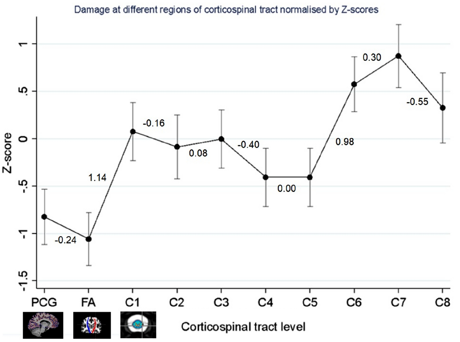
This approach was applied to address competing hypotheses of directionality of neurodegeneration, whether anterograde, cranio-caudal dying-forward from precentral gyrus (posterior frontal lobe of the brain) toward the cervical spinal cord, or dying-back with the disease progressing in the other direction.
In this cross-sectional study, MRI was performed on 75 Motor neuron disease patients and 13 healthy controls. MRI scans were reviewed by a consultant neuroradiologist to exclude significant confounding pathology.
Precentral gyral thickness was estimated from volumetric T1-weighted images using FreeSurfer, corticospinal tract fractional anisotropy from diffusion tensor imaging using FSL, and cross-sectional cervical cord area between C1-C8 levels using Spinal Cord Toolbox.
To analyse these multimodal data within a common domain, individual parameter estimates representing tissue damage at each corticospinal tract level were first converted to z-scores, referenced to healthy control norms.
Mixed-effects linear regression models were then fitted to these z-scores, with gradients hypothesized to represent directionality of neurodegeneration.
The results are a bit confusing, it seems that from C5, there is a forward propagation toward lower cervical vertebras, while above C1 there is a backward propagation toward precentral gyrus!
A major limitation of this study is that they only studied the brain and the upper part of the spinal cord. Upper motor neurons have their body in the motor area and extend to the junction with lower motor neurons (with eventually inter-neurons in between). The lowest corticospinal nerve they studied C8, contributes to the motor innervation of many of the muscles in the trunk and upper limb. Its primary function is the flexion of the fingers. So this study would be conclusive only if all patients had a bulbar pathology, yet only 17 patients on 75 had a bulbar onset.