Today we will present a BioRxiv preprint which has an indirect but interesting link to neurodegenerescence.
By using wavelet transform analysis Sánchez-Moguel et al report that the elderly with excess in theta activity show a higher energy in delta, theta, and alpha bands during the categorization of stimuli in a counting Stroop task.
 Source Vanderbit.edu/Daniel Levin
Their findings imply that this increase neuronal activity might be related to a dysregulated energy metabolism in the elderly with theta excess that could explain the progress to cognitive impairment in this group.
The wavelet analysis allows them to have a standard frequency decomposition of EEG signals over time. So it can help to know the amount of energy used during the execution of Stroop tasks.
 Source WIkipedia/Gerhard Rigoll
Healthy aging is accompanied by a natural detriment of physical and cognitive abilities. In particular, inhibitory control and attention are affected. The general objective of this study was to explore, using Wavelet transform, if there were differences in the amount of energy during the performance of a counting Stroop task between a group of elderly with an excess of theta activity in their EEG and a group of elderly with normal EEG. The specific objective was to evaluate the amount of energy between EEG groups for each of the frequency bands (i.e., delta, theta, alpha, beta, and gamma) across different time windows.
The researchers expected to find higher energy in the group with theta excess, specifically in the time window associated with categorization of stimuli. Changes in brain electrical activity, which can be measured noninvasively by the EEG, are tightly related to cognitive processes. Some authors have proposed that changes in the EEG of the elderly, obtained under resting conditions, are not only the result of normal aging but can contain signs of undergoing subclinical pathologic processe.
Moreover, excess in delta and theta band frequencies of resting EEG from healthy elderly, compared to a normative base according to age, is an excellent predictor of cognitive detriment in the following seven years.
Stroop tasks have been used during functional magnetic resonance imaging (fMRI) to study the decrease in the efficiency of inhibitory processing during healthy and pathological aging. In the counting Stroop task, subjects are asked to answer how many words are presented in a slide, regardless of the meaning of the word itself. Subjects increase their response times and tend to make more mistakes when the meaning of the word does not match the number of times that the word appears; this phenomenon is known as the Stroop or interference effect.
An over-recruitment of neuronal activity during aging was observed using fMRI during the execution of Stroop tasks; this enhanced neuronal activity is proposed to have a compensatory function. Furthermore, fMRI studies showed higher brain activity in older people with mild cognitive impairment compared to healthy elderly.
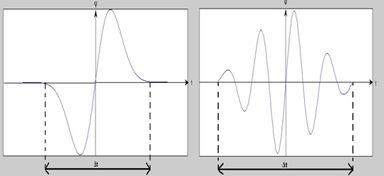
The main advantage of wavelet analysis over Fourier analysis is that they can follow the brain frequency dynamics over time.
The energy analysis using wavelets showed that during the execution of the Stroop task 1. Theta-EEG group assigns a greater amount of total energy in delta, theta, and alpha bands than the Normal-EEG group. 2. Theta-EEG group demands a higher amount of relative energy in delta band but less energy in beta and gamma bands than the normal-EEG group. 3. Theta-EEG group uses higher total energy in all-time windows in the delta, theta, alpha, and beta bands. 4. In the theta and alpha bands, the energy is greater in the Theta-EEG group.
The researchers propose that this excessive energy expenditure in the Theta-EEG group is due because more neurons are recruited in order to perform the task with the same efficiency as the Normal-EEG group. However, they do not know if this energy expenditure is due to cellular and metabolic imbalances that promote progress to cognitive impairment.
Imaging techniques such as fMRI, diffusion tensor imaging, and magnetic resonance spectroscopy, that evaluate the neural networks involved in the task and metabolic expenditure, would complement the findings of the article authors.