Several studies have demonstrated diverse pathological effects of ALS' cerebrospinal fluid in different in vivo and in vitro models. The cerebrospinal fluid is very roughly similar to the lymphatic system, as it drains waste products from the brain metabolism and they are removed into the bloodstream as CSF is absorbed.
Recently an article by Jean-Pierre Julien's group was published on transmission of ALS pathogenesis by the cerebrospinal fluid and we reported it here.
This new article, from Stefan Bräuer and al, is both similar and different to those other articles. On one hand the German scientists from Dresden and Rostock tell that ALS CSF degrades motor neurons, but on the other hand it did report of proteinopathies. It is also a study done in dish versus studies done on living animals.
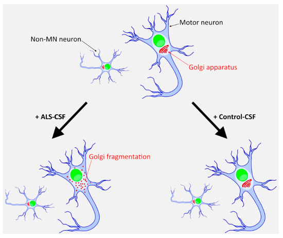
In other studies a multitude of phenotypes linked to ASL-CSF had been observed, including aggregation of transactive response DNA binding protein 43 kDA (TDP-43) in the case of ALS-frontotemporal dementia (FTLD)-CSF, neurofilament abnormalities, gliosis, endoplasmic reticulum (ER)-stress, mitochondrial dysfunction and Golgi fragmentation.
Interesting is the fact that ALS-CSF is able to induce Golgi fragmentation and the inhibition of ER-Golgi trafficking. But this study was not able to induce any proteopathy.
The term Golgi fragmentation describes the dispersion of this cisternal structure across the cell. Golgi fragmentation in motoneurons themselves has been reported in different in vitro and in vivo ALS models. It is also present in postmortem sections of ALS patients.
Moreover, Golgi fragmentation is reported in several other neurodegenerative diseases like Alzheimer’s disease and Parkinson’s disease. There is convincing evidence that Golgi fragmentation is an early event in the disease process and not only a consequence of apoptosis but rather inducing it.
Thus, similar to other neurodegenerative disease conditions, Golgi fragmentation in ALS could be an event that takes place before seeding of the aggregates, axon degeneration and manifestation of clinical symptoms occurs.
This new study aimed at learning how ALS-CSF derived from sporadic ALS patients, induces motor neuronal degeneration in human patient-derived induced pluripotent stem cell (iPSC)-derived motoneurons. Only one aspect of ALS (namely a model for spinal cord degeneration), and thus there might be differences when analyzing other neuronal cell populations affected, e.g., cortical neurons.
CSF was collected from 19 patients. The ALS group consisted of 11 patients (3 women and 8 men) with a mean age of 63 years. The group of 8 control patients (3 women and 5 men) had a mean age of 47 years.
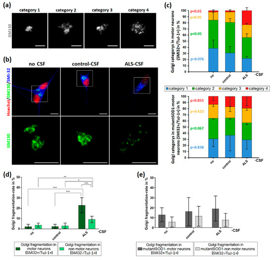
The authors did not observe significant neuronal network degeneration, motoneurons-loss or protein aggregates containing SOD1, FUS or TDP-43, which is a bit surprising Since Golgi fragmentation is expected to be an early event in the pathophysiological cascade, it is plausible that it occurs prior to further changes, which could develop either later or under additional stress conditions.
Additionally they did not observed pathological protein mislocalization or aggregate formation when treating monogenetic ALS-patient-derived motoneurons with ALS-CSF
A major concern regards the cellular age. Because iPSC-derived neurons do resemble fetal neurons and are not as old as the ones normally affected in ALS patients, human development takes much longer.
This study does not explain what is the pathologic agent in the CSF. However it is another study that hints that the etiology of ALS is of extracellular origin.
